File:Pterosaur respiratory system.jpg
Vai alla navigazione
Vai alla ricerca

Dimensioni di questa anteprima: 444 × 599 pixel. Altre risoluzioni: 178 × 240 pixel | 356 × 480 pixel | 569 × 768 pixel | 759 × 1 024 pixel | 1 517 × 2 048 pixel | 3 006 × 4 057 pixel.
File originale (3 006 × 4 057 pixel, dimensione del file: 2,52 MB, tipo MIME: image/jpeg)
| Questo file e la sua pagina di descrizione (discussione · modifica) si trovano su Wikimedia Commons (?) |
| Descrizione |
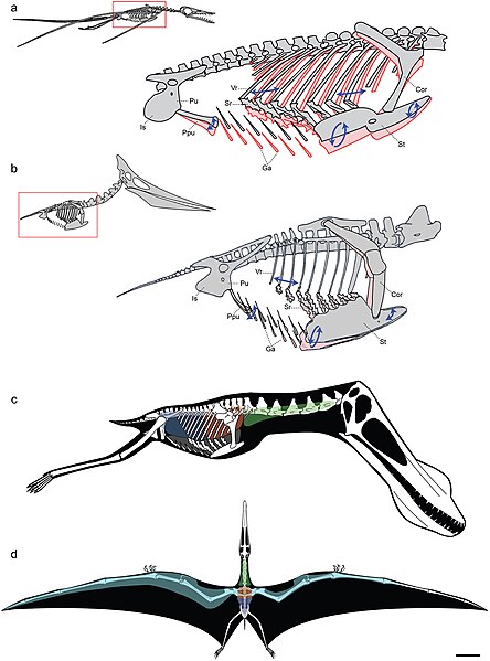
Models of ventilatory kinematics and the pulmonary air sac system of pterosaurs. a, Model of ventilatory kinematics in Rhamphorhynchus. Thoracic movement induced by the ventral intercostal musculature results in forward and outward displacement of the distal vertebral and proximal sternal ribs, and ventral displacement of the sternum, upon inspiration (blue arrows and pink outline). In addition, ventral expansion of the abdomen is induced through caudoventral rotation of the prepubis. Ranges of skeletal movement were modelled after those observed in vivo in the avian thorax and the crocodylian pelvis [26], [27]. Rhamphorhynchus modified from Wellnhofer [48]. b, Model of ventilatory kinematics in Pteranodon wherein the fused anterior vertebral ribs and articulation of the scapulocoracoid with the supraneural plate and anterior sternum limit movement of the anterior sternum, which cannot undergo elliptical rotation. However, the posterior vertebral ribs, sternal ribs, sternum, and prepubis are still capable of anterodorsal-posteroventral excursions facilitating volumetric increases and decreases of the thorax during inspiration-expiration. Pteranodon modified from Bennett [29]. c, d, reconstruction of pulmonary air sac system in the Lower Cretaceous ornithocheirid Anhanguera santanae (AMNH 22555). c, Lateral view showing the inferred position of the lungs (orange), cervical (green) and abdominal air sacs (blue), as predicted on the basis of postcranial skeletal pneumaticity. Thoracic air sacs (shown in grey) are also likely to have been present, but generally do not leave a distinct osteological trace. Humerus and more distal forelimb not shown. d, Dorsal view illustrating the inferred position of subcutaneous diverticular networks (light blue) distally along the wing. The right side depicts a conservative estimate for the size of the airsac network, limiting it to the pre-axial margin of the wing based solely on the presence of pneumatic foramina in closely positioned wing bones. The left side depicts the likely maximal size of an inferred diverticular network, accounting for its inclusion between the dorsal and ventral layers of the wing membrane. Scale = 10 cm. Skeletal reconstruction in c, d modified from Wellnhofer [49]. Abbreviations: as in figure 2, and: Cor: coracoid portion of scapulocoracoid, Ga: gastralia. |
| Data | |
| Fonte | http://www.plosone.org/article/info%3Adoi%2F10.1371%2Fjournal.pone.0004497;jsessionid=A57F0FDB595AC49992E2B5A390FA104C |
| Autore | Leon P. A. M. Claessens, Patrick M. O'Connor, David M. Unwin |
|
Questo file è disponibile in base alla licenza Creative Commons Attribuzione 2.5 Generico
|
Questo file è stato pubblicato in un periodico della Public Library of Science. Il loro sito web afferma che il contenuto dei periodici della PLOS è pubblicato con licenza Creative Commons Attribuzione 4.0 (precedentemente era usata la licenza Creative Commons Attribuzione 2.5), a meno che sia indicato diversamente.
|
Cronologia del file
Fare clic su un gruppo data/ora per vedere il file come si presentava nel momento indicato.
| Data/Ora | Miniatura | Dimensioni | Utente | Commento | |
|---|---|---|---|---|---|
| attuale | 23:17, 2 mar 2009 | | 3 006 × 4 057 (2,52 MB) | FunkMonk | {{Information |Description=Models of ventilatory kinematics and the pulmonary air sac system of pterosaurs. a, Model of ventilatory kinematics in Rhamphorhynchus. Thoracic movement induced by the ventral intercostal musculature results in forward and out |
Pagine che usano questo file
La seguente pagina usa questo file:
Utilizzo globale del file
Anche i seguenti wiki usano questo file:
- Usato nelle seguenti pagine di ar.wikipedia.org:
- Usato nelle seguenti pagine di en.wikipedia.org:
- Usato nelle seguenti pagine di es.wikipedia.org:
- Usato nelle seguenti pagine di ko.wikipedia.org:
- Usato nelle seguenti pagine di nl.wikipedia.org:
- Usato nelle seguenti pagine di oc.wikipedia.org:
- Usato nelle seguenti pagine di outreach.wikimedia.org:
- Usato nelle seguenti pagine di pt.wikipedia.org:
- Usato nelle seguenti pagine di ru.wikipedia.org:
- Usato nelle seguenti pagine di tr.wikipedia.org:
- Usato nelle seguenti pagine di vi.wikipedia.org:
- Usato nelle seguenti pagine di zh.wikipedia.org: