File:Metastasis sites for common cancers.svg

File originale (file in formato SVG, dimensioni nominali 680 × 1 090 pixel, dimensione del file: 1,64 MB)
| Questo file e la sua pagina di descrizione (discussione · modifica) si trovano su Wikimedia Commons (?) |
Dettagli
| Descrizione |
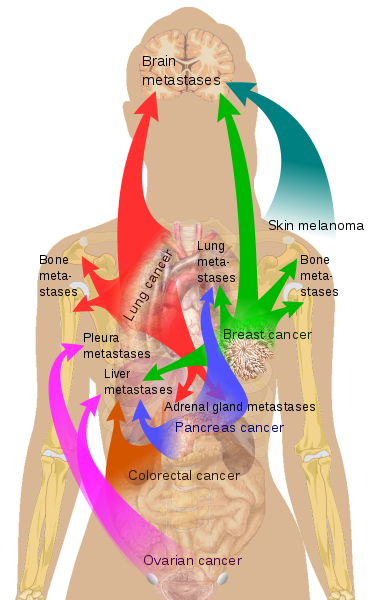
English: Main sites of metastases for common cancer types. Primary cancers are denoted by "...cancer" (except for skin melanoma) and their main metastasis sites are denoted by "...metastases".
List of included informationThe included cancer types are the ones causing most death as per data from the US in 2008.[1]
Not included information of major importance
References
|
| Data | |
| Fonte | All imaged used are in the Public Domain |
| Autore | Mikael Häggström |
| Altre versioni |
[]
|


Licenza
| Questo file è reso disponibile nei termini della licenza Creative Commons CC0 1.0 Universal. | |
| La persona che ha associato un'opera con questo atto legale ha donato tale opera nel pubblico dominio rinunciando a tutti i diritti sull'opera in tutto il mondo, inclusi tutti i diritti connessi o altri diritti simili, per quanto permesso dalla legge. Puoi copiare, modificare, distribuire ed utilizzare l'opera, anche a fini commerciali, senza chiedere alcun permesso.
|
Human body diagramsMain article at: Human body diagrams Template location:Template:Human body diagrams How to derive an imageDerive directly from raster image with organsThe raster (.png format) images below have most commonly used organs already included, and text and lines can be added in almost any graphics editor. This is the easiest method, but does not leave any room for customizing what organs are shown. Adding text and lines: Derive "from scratch"By this method, body diagrams can be derived by pasting organs into one of the "plain" body images shown below. This method requires a graphics editor that can handle transparent images, in order to avoid white squares around the organs when pasting onto the body image. Pictures of organs are found on the project's main page. These were originally adapted to fit the male shadow/silhouette.
Organs:
Derive by vector templateThe Vector templates below can be used to derive images with, for example, Inkscape. This is the method with the greatest potential. See Human body diagrams/Inkscape tutorial for a basic description in how to do this.
Examples of derived works
Licensing
|


.png)


Cronologia del file
Fare clic su un gruppo data/ora per vedere il file come si presentava nel momento indicato.
| Data/Ora | Miniatura | Dimensioni | Utente | Commento | |
|---|---|---|---|---|---|
| attuale | 12:53, 19 ott 2018 | | 680 × 1 090 (1,64 MB) | Jmarchn | Draw now with 3 layers (draw, arrows and text) |
| 09:17, 17 giu 2011 |  | 680 × 1 090 (1,65 MB) | Mikael Häggström | Retry | |
| 07:02, 23 mag 2011 |  | 680 × 1 090 (1,65 MB) | Mikael Häggström | Bunny-jumping, because it won't accept my latest edit. It's always the previous version that shows correct | |
| 07:00, 23 mag 2011 |  | 621 × 767 (1,18 MB) | Mikael Häggström | see above | |
| 06:56, 23 mag 2011 |  | 680 × 1 090 (1,65 MB) | Mikael Häggström | Now it's to previous one that shows correct! Reverting. | |
| 06:55, 23 mag 2011 |  | 680 × 1 090 (1,65 MB) | Mikael Häggström | No response, so redid | |
| 06:54, 23 mag 2011 |  | 680 × 1 090 (1,65 MB) | Mikael Häggström | Made pancreas arrows blue to distinguish from the red of lung cancer | |
| 18:12, 22 mag 2011 |  | 680 × 1 090 (1,65 MB) | Mikael Häggström | darker green | |
| 17:57, 22 mag 2011 |  | 680 × 1 090 (1,65 MB) | Mikael Häggström | Ups, forgot to write out colorectal cancer | |
| 17:55, 22 mag 2011 |  | 680 × 1 090 (1,65 MB) | Mikael Häggström | {{Information |Description ={{en|1=f}} |Source ={{own}} |Author =Mikael Häggström |Date =f |Permission = |other_versions = }} |
Pagine che usano questo file
Nessuna pagina utilizza questo file.
Utilizzo globale del file
Anche i seguenti wiki usano questo file:
- Usato nelle seguenti pagine di ar.wikipedia.org:
- Usato nelle seguenti pagine di en.wikipedia.org:
- Usato nelle seguenti pagine di eu.wikipedia.org:
- Usato nelle seguenti pagine di fa.wikipedia.org:
- Usato nelle seguenti pagine di ha.wikipedia.org:
- Usato nelle seguenti pagine di hy.wikipedia.org:
- Usato nelle seguenti pagine di sr.wikipedia.org:
- Usato nelle seguenti pagine di tl.wikipedia.org:
- Usato nelle seguenti pagine di zh.wikipedia.org: