File:Echinococcus Life Cycle.svg
Vai alla navigazione
Vai alla ricerca

Dimensioni di questa anteprima PNG per questo file SVG: 629 × 600 pixel. Altre risoluzioni: 252 × 240 pixel | 504 × 480 pixel | 806 × 768 pixel | 1 074 × 1 024 pixel | 2 149 × 2 048 pixel | 1 280 × 1 220 pixel.
File originale (file in formato SVG, dimensioni nominali 1 280 × 1 220 pixel, dimensione del file: 643 KB)
| Questo file e la sua pagina di descrizione (discussione · modifica) si trovano su Wikimedia Commons (?) |
Dettagli
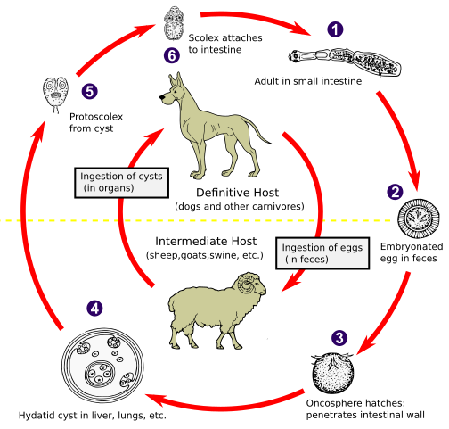
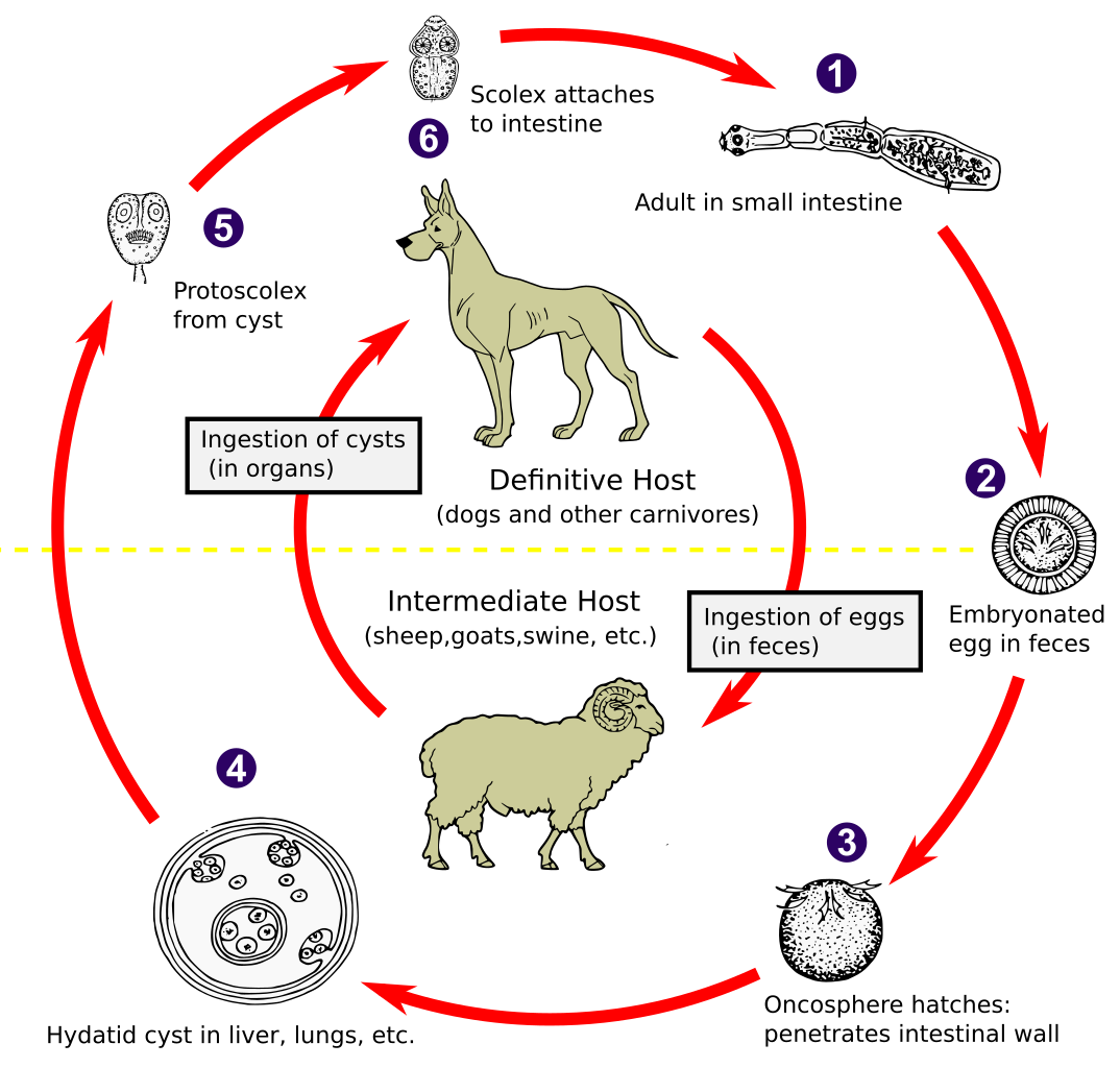
| Descrizione | The adult Echinococcus granulosus (3 to 6 mm long) [1] resides in the small bowel of the definitive hosts (dogs or other carnivores). Gravid proglottids release eggs [2] that are passed in the feces. After ingestion by a suitable intermediate host (under natural conditions: sheep, goat, swine, cattle, horses, camel), the egg hatches in the small bowel and releases an oncosphere [3] that penetrates the intestinal wall and migrates through the circulatory system into various organs, especially the liver and lungs. In these organs, the oncosphere develops into a cyst [4] that enlarges gradually, producing protoscolices and daughter cysts that fill the cyst interior. The definitive host becomes infected by ingesting the cyst-containing organs of the infected intermediate host. After ingestion, the protoscolices [5] evaginate, attach to the intestinal mucosa [6] and develop into adult stages [1] in 32 to 80 days. The same life cycle occurs with E. multilocularis (1.2 to 3.7 mm), with the following differences: the definitive hosts are foxes, and to a lesser extent dogs, cats, coyotes and wolves; the intermediate host are small rodents; and larval growth (in the liver) remains indefinitely in the proliferative stage, resulting in invasion of the surrounding tissues. With E. vogeli (up to 5.6 mm long), the definitive hosts are bush dogs and dogs; the intermediate hosts are rodents; and the larval stage (in the liver, lungs and other organs) develops both externally and internally, resulting in multiple vesicles. E. oligarthrus (up to 2.9 mm long) has a life cycle that involves wild felids as definitive hosts and rodents as intermediate hosts. Humans become infected by ingesting eggs , with resulting release of oncospheres in the intestine and the development of cysts in various organs. Image adapted from original available at the United States Centres for Disease Control Parasitology Identification Laboratory ([1]). |
| Data | |
| Fonte |
Questo file deriva da: Echinococcus Life Cycle.png: |
| Autore |
CDC Vettoriale: 🎱 |
| SVG sviluppo | Il codice sorgente di questo file SVG non è valido a causa di 2 errori. Questo diagramma in grafica vettoriale non valido secondo il W3C è stato creato con un programma SVG ignoto. |
Licenza
This image is a work of the Centers for Disease Control and Prevention, part of the United States Department of Health and Human Services, taken or made as part of an employee's official duties. As a work of the U.S. federal government, the image is in the public domain.
|
Registro originale del caricamento
This image is a derivative work of the following images:
- Echinococcus Life Cycle.png licensed with PD-USGov-HHS-CDC
- 2007-01-24T10:54:56Z Pngbot 600x571 (45555 Bytes) optimized with optipng
- 2005-04-26T01:48:50Z FirstPrinciples~commonswiki 600x571 (55999 Bytes) Smaller & clearer
- 2005-04-26T01:36:23Z FirstPrinciples~commonswiki 800x761 (80990 Bytes)
Uploaded with derivativeFX
Cronologia del file
Fare clic su un gruppo data/ora per vedere il file come si presentava nel momento indicato.
| Data/Ora | Miniatura | Dimensioni | Utente | Commento | |
|---|---|---|---|---|---|
| attuale | 03:31, 1 feb 2021 | | 1 280 × 1 220 (643 KB) | Pixelsquid | Resized. |
| 22:44, 31 gen 2021 |  | 320 × 305 (460 KB) | Pixelsquid | == {{int:filedesc}} == {{Information |Description=The adult Echinococcus granulosus (3 to 6 mm long) [1] resides in the small bowel of the definitive hosts (dogs or other carnivores). Gravid proglottids release eggs [2] that are passed in the feces. After ingestion by a suitable intermediate host (under natural conditions: sheep, goat, swine, cattle, horses, camel), the egg hatches in the small bowel and releases an oncosphere [3] that penetrates the intestinal wall and migrates through the... |
Pagine che usano questo file
La seguente pagina usa questo file:
Utilizzo globale del file
Anche i seguenti wiki usano questo file:
- Usato nelle seguenti pagine di ar.wikipedia.org:
- Usato nelle seguenti pagine di arz.wikipedia.org:
- Usato nelle seguenti pagine di be.wikipedia.org:
- Usato nelle seguenti pagine di bs.wikipedia.org:
- Usato nelle seguenti pagine di ca.wikipedia.org:
- Usato nelle seguenti pagine di dag.wikipedia.org:
- Usato nelle seguenti pagine di el.wikipedia.org:
- Usato nelle seguenti pagine di en.wikipedia.org:
- Usato nelle seguenti pagine di es.wikipedia.org:
- Usato nelle seguenti pagine di fa.wikipedia.org:
- Usato nelle seguenti pagine di ga.wikipedia.org:
- Usato nelle seguenti pagine di gl.wikipedia.org:
- Usato nelle seguenti pagine di hi.wikipedia.org:
- Usato nelle seguenti pagine di hu.wikipedia.org:
- Usato nelle seguenti pagine di hy.wikipedia.org:
- Usato nelle seguenti pagine di ia.wikipedia.org:
- Usato nelle seguenti pagine di id.wikipedia.org:
- Usato nelle seguenti pagine di is.wikipedia.org:
- Usato nelle seguenti pagine di ja.wikipedia.org:
- Usato nelle seguenti pagine di ko.wikipedia.org:
- Usato nelle seguenti pagine di ky.wikipedia.org:
- Usato nelle seguenti pagine di lt.wikipedia.org:
- Usato nelle seguenti pagine di mk.wikipedia.org:
- Usato nelle seguenti pagine di ml.wikipedia.org:
- Usato nelle seguenti pagine di ms.wikipedia.org:
- Usato nelle seguenti pagine di nl.wikipedia.org:
- Usato nelle seguenti pagine di om.wikipedia.org:
- Usato nelle seguenti pagine di or.wikipedia.org:
- Usato nelle seguenti pagine di pl.wikipedia.org:
- Usato nelle seguenti pagine di pt.wikipedia.org:
- Usato nelle seguenti pagine di ro.wikipedia.org:
- Usato nelle seguenti pagine di ru.wikipedia.org:
- Usato nelle seguenti pagine di sl.wikipedia.org:
- Usato nelle seguenti pagine di sr.wikipedia.org:
- Usato nelle seguenti pagine di sv.wikipedia.org:
- Usato nelle seguenti pagine di th.wikipedia.org:
- Usato nelle seguenti pagine di tl.wikipedia.org:
- Usato nelle seguenti pagine di tr.wikipedia.org:
- Usato nelle seguenti pagine di uk.wikipedia.org:
- Usato nelle seguenti pagine di uz.wikipedia.org:
- Usato nelle seguenti pagine di vi.wikipedia.org:
- Usato nelle seguenti pagine di www.wikidata.org:
Visualizza l'utilizzo globale di questo file.