File:Anaplastic astrocytoma.jpg
Vai alla navigazione
Vai alla ricerca
Dimensioni di questa anteprima: 800 × 552 pixel. Altre risoluzioni: 320 × 221 pixel | 640 × 442 pixel | 1 024 × 707 pixel | 1 200 × 828 pixel.
File originale (1 200 × 828 pixel, dimensione del file: 200 KB, tipo MIME: image/jpeg)
| Questo file e la sua pagina di descrizione (discussione · modifica) si trovano su Wikimedia Commons (?) |
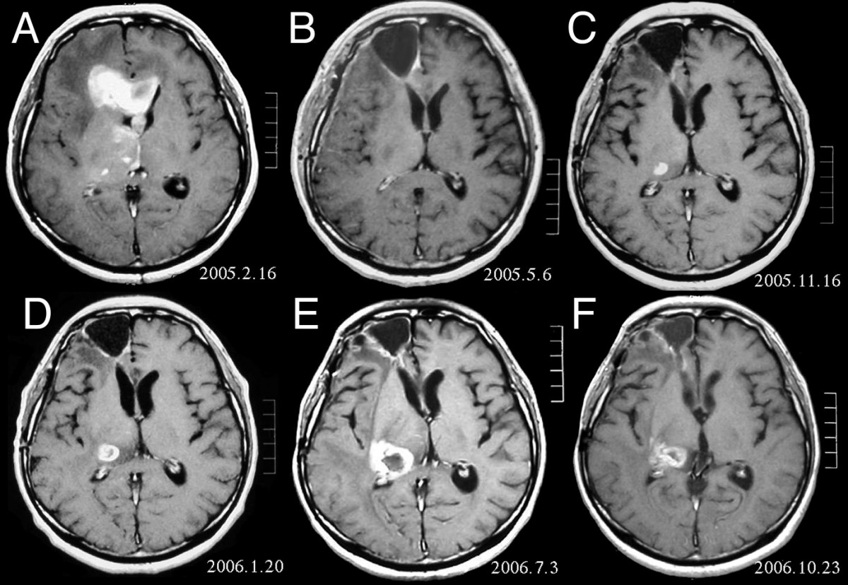
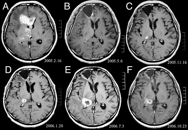
| Descrizione | MRI of brain. (A) Initial MRI on February 16, 2005, shows a tumor in the right and left frontal lobe as well as the right thalamus. (B) MRI after surgery, radiation and chemotherapy. The tumor has completely disappeared except for slight enhancement adjacent to the surgical margin. (C) Recurrence of the thalamic tumor despite maintenance chemotherapy on November 16, 2005. (D) Increase in size of the thalamic tumor two months after stereotactic radiotherapy. (E) After 6 cycles of TMZ therapy, the thalamic lesion enlarged, and the patient developed dysarthria and hemiparesis. (F) After 2 courses of treatment with interferon-beta and TMZ, the tumor shows a partial response. |
| Data | |
| Fonte | Fujimaki T, Ishii H, Matsuno A, Arai H, Nakagomi T.Effectiveness of interferon-beta and temozolomide combination therapy against temozolomide-refractory recurrent anaplastic astrocytoma.World J Surg Oncol. 2007 Aug 4;5:89. PMID 17683572 doi:10.1186/1477-7819-5-89 |
| Autore | Fujimaki T, Ishii H, Matsuno A, Arai H, Nakagomi T. |
| Licenza (Riusare questo file) |
BioMedCentral License |
Questo file è disponibile in base alla licenza Creative Commons Attribuzione 2.0 Generico
- Tu sei libero:
- di condividere – di copiare, distribuire e trasmettere quest'opera
- di modificare – di adattare l'opera
- Alle seguenti condizioni:
- attribuzione – Devi fornire i crediti appropriati, un collegamento alla licenza e indicare se sono state apportate modifiche. Puoi farlo in qualsiasi modo ragionevole, ma non in alcun modo che suggerisca che il licenziante approvi te o il tuo uso.
Cronologia del file
Fare clic su un gruppo data/ora per vedere il file come si presentava nel momento indicato.
| Data/Ora | Miniatura | Dimensioni | Utente | Commento | |
|---|---|---|---|---|---|
| attuale | 18:47, 25 feb 2008 | | 1 200 × 828 (200 KB) | Filip em | {{Information |Description=MRI of brain. (A) Initial MRI on February 16, 2005, shows a tumor in the right and left frontal lobe as well as the right thalamus. (B) MRI after surgery, radiation and chemotherapy. The tumor has completely disappeared except f |
Pagine che usano questo file
Le seguenti 4 pagine usano questo file:
Utilizzo globale del file
Anche i seguenti wiki usano questo file:
- Usato nelle seguenti pagine di ar.wikipedia.org:
- Usato nelle seguenti pagine di bg.wikipedia.org:
- Usato nelle seguenti pagine di cs.wikipedia.org:
- Usato nelle seguenti pagine di da.wikipedia.org:
- Usato nelle seguenti pagine di de.wikipedia.org:
- Usato nelle seguenti pagine di el.wikipedia.org:
- Usato nelle seguenti pagine di en.wikipedia.org:
- Usato nelle seguenti pagine di es.wikipedia.org:
- Usato nelle seguenti pagine di et.wikipedia.org:
- Usato nelle seguenti pagine di hi.wikipedia.org:
- Usato nelle seguenti pagine di hr.wikipedia.org:
- Usato nelle seguenti pagine di hu.wikipedia.org:
- Usato nelle seguenti pagine di kk.wikipedia.org:
- Usato nelle seguenti pagine di lb.wikipedia.org:
- Usato nelle seguenti pagine di lv.wikipedia.org:
- Usato nelle seguenti pagine di mk.wikipedia.org:
- Usato nelle seguenti pagine di mt.wikipedia.org:
- Usato nelle seguenti pagine di nl.wikipedia.org:
- Usato nelle seguenti pagine di no.wikipedia.org:
- Usato nelle seguenti pagine di pl.wikipedia.org:
- Usato nelle seguenti pagine di ro.wikipedia.org:
- Usato nelle seguenti pagine di sk.wikipedia.org:
- Usato nelle seguenti pagine di sl.wikipedia.org:
- Usato nelle seguenti pagine di sq.wikipedia.org:
- Usato nelle seguenti pagine di sr.wikipedia.org:
- Usato nelle seguenti pagine di uk.wikipedia.org: