Area 9 di Brodmann
| Area 9 di Brodmann | |
|---|---|
 | |
 | |
| Nome latino | Area frontalis granularis |
| Sistema | Sistema nervoso centrale |
| Identificatori | |
| FMA | 68606 |
| ID NeuroLex | birnlex_1740 |
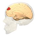
L' area 9 di Brodmann, nell'acronimo inglese BA9, è un'area della corteccia frontale del cervello umano e di altri primati. Contribuisce al funzionamento della corteccia dorsolaterale prefrontale e della corteccia prefrontale mediale.
Scimmia[modifica | modifica wikitesto]
Il termine area 9 di Brodmann si riferisce a una porzione del lobo frontale del cercopithecus definita tramite la citoarchitettonica. Brodmann-1909 la considerava topograficamente e citoarchitettonicamente omologa alla corteccia granulare frontale (area 9) e all'area frontopolare (area 10) degli umani.
Le caratteristiche distintive rispetto alle altre aree sono molteplici (Brodmann-1905): l'area nove, diversamente dall'area 6, ha uno strato granulare interno (IV) definito; diversamente dall'area 6 e l'area 8, il suo strato piramidale interno (V) è divisibile in due substrati, uno strato 5a che si fonde parzialmente con lo strato IV, contenente una grande densità di cellule gangliari di media grandezza, e uno strato 5b più interno, chiaro e povero di cellule; le cellule piramidali del substrato 3b dello strato piramidale esterno (III) sono piccole e sparse; lo strato granulare esterno (II) è stretto e contiene poche cellule granulari sparse.[1]
Funzioni[modifica | modifica wikitesto]
L'area è coinvolta nella gestione della memoria a breve termine,[2] la valutazione delle attività recenti,[3] le risposte automatiche,[4] la fluidità verbale,[5] il rilevamento degli errori,[6] l'attenzione uditiva verbale,[7] la comprensione delle intenzioni altrui,[8] la comprensione delle immagini spaziali,[9] il ragionamento induttivo,[10] l'attribuzione delle intenzioni,[11] nel mantenere alta l'attenzione durante il conteggio di stimoli uditivi,[12] e nel più basso consumo di energia negli individui con disturbi bipolari.[13]
L'area che si trova nell'emisfero sinistro è parzialmente responsabile dell'empatia[14], nella comprensione degli idiomi,[15][16] nel processare le scene piacevoli e spiacevoli,[17] nell'autocritica,[18] e nel controllo delle emozioni negative.[19]
Nell'emisfero destro la regione è coinvolta nell'attribuzione delle intenzioni,[20] la teoria mentale,[21] la soppressione della tristezza,[22] la memoria di lavoro,[23][24] la memoria spaziale,[25][26] il riconoscimento,[27][28][29] il richiamo,[28][30][31], il riconoscimento delle emozioni degli altri,[32] la pianificazione,[33] il calcolo,[34][35] l'elaborazione semantica e percettiva degli odori,[36] la religiosità,[37] e l'attenzione alle emozioni positive.[19]
Galleria d'immagini[modifica | modifica wikitesto]
-
 Animazione
 -
 Vista frontale
 -
 Vista laterale
 -
 Vista mediale


Note[modifica | modifica wikitesto]
- ^ BrainInfo, su braininfo.rprc.washington.edu. URL consultato il 9 dicembre 2015.
- ^ Claudio Babiloni, Antonio Ferretti e Cosimo Del Gratta, Human cortical responses during one-bit delayed-response tasks: An fMRI study, in Brain Research Bulletin, vol. 65, n. 5, 15 maggio 2005, pp. 383-390, DOI:10.1016/j.brainresbull.2005.01.013. URL consultato il 9 dicembre 2015.
- ^ Wolters Kluwer Health - Article Landing Page, DOI:10.1097/00001756-199611040-00079. URL consultato il 9 dicembre 2015.
- ^ Andrea Kübler, Veronica Dixon e Hugh Garavan, Automaticity and Reestablishment of Executive Control—An fMRI Study, in Journal of Cognitive Neuroscience, vol. 18, n. 8, 21 luglio 2006, pp. 1331-1342, DOI:10.1162/jocn.2006.18.8.1331. URL consultato il 9 dicembre 2015.
- ^ (EN) Sharon Abrahams, Laura H. Goldstein e Andy Simmons, Functional magnetic resonance imaging of verbal fluency and confrontation naming using compressed image acquisition to permit overt responses, in Human Brain Mapping, vol. 20, n. 1, 1º settembre 2003, pp. 29-40, DOI:10.1002/hbm.10126. URL consultato il 9 dicembre 2015.
- ^ (EN) Andre D. Chevrier, Michael D. Noseworthy e Russell Schachar, Dissociation of response inhibition and performance monitoring in the stop signal task using event-related fMRI, in Human Brain Mapping, vol. 28, n. 12, 1º dicembre 2007, pp. 1347-1358, DOI:10.1002/hbm.20355. URL consultato il 9 dicembre 2015.
- ^ Toshiharu Nakai, Chikako Kato e Kayako Matsuo, An fMRI Study to Investigate Auditory Attention: A Model of the Cocktail Party Phenomenon, in Magnetic Resonance in Medical Sciences, vol. 4, n. 2, 1º gennaio 2005, pp. 75-82, DOI:10.2463/mrms.4.75. URL consultato il 9 dicembre 2015.
- ^ Vinod Goel, Jordan Grafman e Norihiro Sadato, Modeling other minds, in NeuroReport, vol. 6, n. 13, pp. 1741-1746, DOI:10.1097/00001756-199509000-00009. URL consultato il 9 dicembre 2015.
- ^ Markus Knauff, Thomas Mulack e Jan Kassubek, Spatial imagery in deductive reasoning: a functional MRI study, in Cognitive Brain Research, vol. 13, n. 2, 1º aprile 2002, pp. 203-212, DOI:10.1016/S0926-6410(01)00116-1. URL consultato il 9 dicembre 2015.
- ^ Vinod Goel, Brian Gold e Shitij Kapur, The seats of reason? An imaging study of deductive and inductive reasoning, in NeuroReport, vol. 8, n. 5, pp. 1305-1310, DOI:10.1097/00001756-199703240-00049. URL consultato il 9 dicembre 2015.
- ^ (EN) Gereon R. Fink, John C. Marshall e Peter W. Halligan, The neural consequences of conflict between intention and the senses, in Brain, vol. 122, n. 3, 1º marzo 1999, pp. 497-512, DOI:10.1093/brain/122.3.497. URL consultato il 9 dicembre 2015.
- ^ Tim Shallice, Donald T. Stuss e Michael P. Alexander, The multiple dimensions of sustained attention, in Cortex, vol. 44, n. 7, 1º luglio 2008, pp. 794-805, DOI:10.1016/j.cortex.2007.04.002. URL consultato il 9 dicembre 2015.
- ^ (EN) John O Brooks, Carrie E Bearden e Jennifer C Hoblyn, Prefrontal and paralimbic metabolic dysregulation related to sustained attention in euthymic older adults with bipolar disorder, in Bipolar Disorders, vol. 12, n. 8, 1º dicembre 2010, pp. 866-874, DOI:10.1111/j.1399-5618.2010.00881.x. URL consultato il 9 dicembre 2015.
- ^ Tom F. D. Farrow, Ying Zheng e Iain D. Wilkinson, Investigating the functional anatomy of empathy and forgiveness, in Neuroreport, vol. 12, n. 11, pp. 2433-2438, DOI:10.1097/00001756-200108080-00029. URL consultato il 9 dicembre 2015.
- ^ Richard J. Maddock e Michael H. Buonocore, Activation of left posterior cingulate gyrus by the auditory presentation ofthreat-related words: an fMRI study, in Psychiatry Research: Neuroimaging, vol. 75, n. 1, pp. 1-14, DOI:10.1016/s0925-4927(97)00018-8. URL consultato il 9 dicembre 2015.
- ^ (EN) Leonor J. Romero Lauro, Marco Tettamanti e Stefano F. Cappa, Idiom Comprehension: A Prefrontal Task?, in Cerebral Cortex, vol. 18, n. 1, 1º gennaio 2008, pp. 162-170, DOI:10.1093/cercor/bhm042. URL consultato il 9 dicembre 2015.
- ^ Richard D. Lane, Eric M. Reiman e Margaret M. Bradley, Neuroanatomical correlates of pleasant and unpleasant emotion, in Neuropsychologia, vol. 35, n. 11, 1º novembre 1997, pp. 1437-1444, DOI:10.1016/S0028-3932(97)00070-5. URL consultato il 9 dicembre 2015.
- ^ Olivia Longe, Frances A. Maratos e Paul Gilbert, Having a word with yourself: Neural correlates of self-criticism and self-reassurance, in NeuroImage, vol. 49, n. 2, 15 gennaio 2010, pp. 1849-1856, DOI:10.1016/j.neuroimage.2009.09.019. URL consultato il 9 dicembre 2015.
- ^ a b R. Kerestes, C. D. Ladouceur e S. Meda, Abnormal prefrontal activity subserving attentional control of emotion in remitted depressed patients during a working memory task with emotional distracters, in Psychological Medicine, vol. 42, n. 01, 1º gennaio 2012, pp. 29–40, DOI:10.1017/S0033291711001097. URL consultato il 9 dicembre 2015.
- ^ Eric Brunet, Yves Sarfati e Marie-Christine Hardy-Baylé, A PET Investigation of the Attribution of Intentions with a Nonverbal Task, in NeuroImage, vol. 11, n. 2, 1º febbraio 2000, pp. 157-166, DOI:10.1006/nimg.1999.0525. URL consultato il 9 dicembre 2015.
- ^ Helen L. Gallagher, Anthony I. Jack e Andreas Roepstorff, Imaging the Intentional Stance in a Competitive Game, in NeuroImage, vol. 16, 3, Part A, 1º luglio 2002, pp. 814-821, DOI:10.1006/nimg.2002.1117. URL consultato il 9 dicembre 2015.
- ^ Simerjit Kaur, Roberto B. Sassi e David Axelson, Cingulate Cortex Anatomical Abnormalities in Children and Adolescents With Bipolar Disorder, in American Journal of Psychiatry, vol. 162, n. 9, 1º settembre 2005, pp. 1637-1643, DOI:10.1176/appi.ajp.162.9.1637. URL consultato il 9 dicembre 2015.
- ^ John X Zhang, Hoi-Chung Leung e Marcia K Johnson, Frontal activations associated with accessing and evaluating information in working memory: an fMRI study, in NeuroImage, vol. 20, n. 3, 1º novembre 2003, pp. 1531-1539, DOI:10.1016/j.neuroimage.2003.07.016. URL consultato il 9 dicembre 2015.
- ^ Carol L. Raye, Marcia K. Johnson e Karen J. Mitchell, Neuroimaging a Single Thought: Dorsolateral PFC Activity Associated with Refreshing Just-Activated Information, in NeuroImage, vol. 15, n. 2, 1º febbraio 2002, pp. 447-453, DOI:10.1006/nimg.2001.0983. URL consultato il 9 dicembre 2015.
- ^ Scott D. Slotnick e Lauren R. Moo, Prefrontal cortex hemispheric specialization for categorical and coordinate visual spatial memory, in Neuropsychologia, vol. 44, n. 9, 1º gennaio 2006, pp. 1560-1568, DOI:10.1016/j.neuropsychologia.2006.01.018. URL consultato il 9 dicembre 2015.
- ^ H.-C. Leung, J. C. Gore e P. S. Goldman-Rakic, Sustained Mnemonic Response in the Human Middle Frontal Gyrus during On-Line Storage of Spatial Memoranda, in Journal of Cognitive Neuroscience, vol. 14, n. 4, 1º maggio 2002, pp. 659-671, DOI:10.1162/08989290260045882. URL consultato il 9 dicembre 2015.
- ^ Charan Ranganath, Marcia K Johnson e Mark D’Esposito, Prefrontal activity associated with working memory and episodic long-term memory, in Neuropsychologia, vol. 41, n. 3, 1º gennaio 2003, pp. 378-389, DOI:10.1016/S0028-3932(02)00169-0. URL consultato il 9 dicembre 2015.
- ^ a b (EN) M. D. Rugg, P. C. Fletcher e C. D. Frith, Differential activation of the prefrontal cortex in successful and unsuccessful memory retrieval, in Brain, vol. 119, n. 6, 1º dicembre 1996, pp. 2073-2083, DOI:10.1093/brain/119.6.2073. URL consultato il 9 dicembre 2015.
- ^ (EN) Endel Tulving, Reza Habib e Lars Nyberg, Positron emission tomography correlations in and beyond medial temporal lobes [collegamento interrotto], in Hippocampus, vol. 9, n. 1, 1º gennaio 1999, pp. 71-82, DOI:10.1002/(SICI)1098-1063(1999)9:13.0.CO;2-F. URL consultato il 9 dicembre 2015.
- ^ (EN) E. Tulving, S. Kapur e H. J. Markowitsch, Neuroanatomical correlates of retrieval in episodic memory: auditory sentence recognition, in Proceedings of the National Academy of Sciences, vol. 91, n. 6, 15 marzo 1994, pp. 2012-2015, DOI:10.1073/pnas.91.6.2012. URL consultato il 9 dicembre 2015 (archiviato dall'url originale il 29 aprile 2017).
- ^ (EN) Emrah Düzel, Terence W. Picton e Roberto Cabeza, Comparative electrophysiological and hemodynamic measures of neural activation during memory-retrieval, in Human Brain Mapping, vol. 13, n. 2, 1º giugno 2001, pp. 104-123, DOI:10.1002/hbm.1028. URL consultato il 9 dicembre 2015.
- ^ (EN) Felix Bermpohl, Alvaro Pascual-Leone e Amir Amedi, Attentional modulation of emotional stimulus processing: An fMRI study using emotional expectancy, in Human Brain Mapping, vol. 27, n. 8, 1º agosto 2006, pp. 662-677, DOI:10.1002/hbm.20209. URL consultato il 9 dicembre 2015.
- ^ (EN) Jon M. Fincham, Cameron S. Carter e Vincent van Veen, Neural mechanisms of planning: A computational analysis using event-related fMRI, in Proceedings of the National Academy of Sciences, vol. 99, n. 5, 5 marzo 2002, pp. 3346-3351, DOI:10.1073/pnas.052703399. URL consultato il 9 dicembre 2015 (archiviato dall'url originale il 3 giugno 2018).
- ^ Sheng Xie, Jiangxi Xiao e Xuexiang Jiang, [The fMRI study of the calculation tasks in normal aged volunteers], in Beijing Da Xue Xue Bao. Yi Xue Ban = Journal of Peking University. Health Sciences, vol. 35, n. 3, 18 giugno 2003, pp. 311-313. URL consultato il 9 dicembre 2015.
- ^ T. C Rickard, S. G Romero e G Basso, The calculating brain: an fMRI study, in Neuropsychologia, vol. 38, n. 3, 1º marzo 2000, pp. 325-335, DOI:10.1016/S0028-3932(99)00068-8. URL consultato il 9 dicembre 2015.
- ^ Jean-P. Royet, Olivier Koenig e Marie-C. Gregoire, Functional Anatomy of Perceptual and Semantic Processing for Odors, in Journal of Cognitive Neuroscience, vol. 11, n. 1, 1º gennaio 1999, pp. 94-109, DOI:10.1162/089892999563166. URL consultato il 9 dicembre 2015.
- ^ (EN) Nina P. Azari, Janpeter Nickel e Gilbert Wunderlich, Neural correlates of religious experience, in European Journal of Neuroscience, vol. 13, n. 8, 1º aprile 2001, pp. 1649-1652, DOI:10.1046/j.0953-816x.2001.01527.x. URL consultato il 9 dicembre 2015.
Voci correlate[modifica | modifica wikitesto]
Altri progetti[modifica | modifica wikitesto]
 Wikimedia Commons contiene immagini o altri file su Area 9 di Brodmann
Collegamenti esterni[modifica | modifica wikitesto]
- http://www.pnas.org/cgi/reprint/98/7/4259.pdf Archiviato il 17 maggio 2008 in Internet Archive.
- Per la neuroanatomia di quest'area vedere BrainInfo Archiviato il 5 novembre 2005 in Wikiwix.