Utente:Anassagora/Sandbox02

Il doppio strato lipidico è una sottile membrana, composta da due foglietti di molecole lipidiche. La membrana plasmatica di quasi tutti gli organismi viventi e di molti virus è composta un doppio strato lipidico, così come le membrane che circondano il nucleo cellulare e gli altri organelli.
Esso forma una barriera che mantiene gli ioni, le proteine e tutte le altre molecole dove sono necessarie e impedisce loro di diffondere in regioni dove non dovrebbero trovarsi. I doppi strati lipidici sono particolarmente adatti a questo scopo in quanto, pur essendo spessi soltanto pochi nanometri, sono impermeabili alla maggior parte delle molecole idrofile (ovvero polari). Per questo risultano altamente impermeabili agli ioni, permettendo alle cellule di regolare le concentrazoni dei sali e il pH, pompando ioni attraverso le loro membrane utilizzando proteine chiamate pompe ioniche
I doppi strati naturali sono solitamente composti per la maggior parte da fosfolipidi, molecole di natura anfipatica, ovvero, che posseggono una testa idrofilica e due code idrofobiche. Quando i fosfolipidi vengono posti in acqua, si dispongono spontaneamente in un foglietto a due strati con tutte le code rivolte verso l'interno e tutte le teste verso l'esterno. Il centro di questo doppio strato non contiene praticamente alcuna molecola di acqua ed esclude molecole solubili in acqua ma non in olio, come gli zuccheri e i sali. Questo processo di assemblaggio è simile alla formazione di goccioline quando si miscela olio in acqua ed è dovuta alla stessa forza, chiamata effetto idrofobico.
Dal momento che i doppi strati lipidici sono abbastanza fragili e sono così sottili da non poter essere visualizzato al microscopio ottico tradizionale, il loro studio è qualcosa di molto complesso. Esperimenti sui doppi strati lipidici richiedono spesso tecniche avanzate quali la microscopia elettronica e la microscopia a forza atomica.
Fosfolipidi con alcuni gruppi di testa possono alterare la chimica di superficie di un doppio strato e possono, ad esempio, marcare una cellula perché questa venga distrutta dal sistema immunitario. Anche le code lipidiche possono influenzare le proprietà della membrana, determinando ad esempio la fase del doppio strato. Questo può adottare uno stato solido a gel a basse temperature ma andare incontro ad una transizione di fase verso uno stato fluido a temperature più elevate. L'impaccamento dei lipidi all'interno della struttura a doppio strato influenza anche le sue proprietà meccaniche, tra cui la sua resistenza allo stiramento e alla distorsione. Molte di queste proprietà sono state studiate mediante l'utilizzo di modelli artificiali di doppi strati lipidici prodotti in laboratorio. Vescicole prodotte da doppi strati modello sono anche state utilizzate in campo clinico per somministrare dei farmaci.
Le membrane biologiche includono solitamente diversi tipi di lipidi, oltre ai fosfolipidi. Uno particolarmente importante è, ad esempio, il colesterolo, che aiuta a rinforzare il doppio strato e diminuisce la sua permeabilità. Il colesterolo aiuta anche a regolare l'attività di certe proteine integrali di membrana. Queste funzionano quando sono incorporate in un doppio strato lipidico. Poiché i doppi strati definiscono i confini della cellula e dei suoi compartimenti, queste proteine di membrana sono coinvolte in molti processi di segnalazione intracellulare e intercellulare. Alcuni tipi di proteine di membrana sono coinvolti nel processo di fusione di due doppi strati lipidici. Questa fusione permette l'unione di due strutture distinte come ad esempio nella fecondazione di un uovo da parte di uno spermatozoo o l'ingresso di un virus in una cellula.

Struttura e organizzazione[modifica | modifica wikitesto]
Strutturalmente, un doppio strato lipidico è una superficie, composta da lipidi, dello spessore di due molecole, disposte in modo tale che le teste idrofile puntano verso l'esterno, da entrambi i lati del doppio strato, mentre le code idrofobiche puntano verso il suo interno. Questa disposizione dorma due "foglietti", che sono ognuno un singolo strato di molecole. I lipidi si autoassemblano in questa struttura a causa dell'effetto idrofobico, che crea interazioni energeticamente sfavorevoli tra le code lipidiche e l'acqua circostante. Per questo motivo, un doppio strato lipidico è tenuto assieme esclusivamente da interazioni intermolecolari che non coinvolgono la formazione di legami covalenti tra le singole molecole.
Un doppio strato lipidico ha alcuni aspetti in comune con una semplice bolla di sapone, sebbene ci siano anche delle importanti differenze. Entrambe le strutture sono composte da due singoli strati di sostanze anfipatiche. La disposizione delle molecole, tuttavia, nella bolla di sapone è esattamente l'opposto di quella del doppio strato lipidico. I due monostrati di sapone, infatti, ricoprono un velo d'acqua situato tra i due. Per questo motivo, le teste sono rivolte verso il centro acquoso, mentre le code sono rivolte verso l'esterno. Un'altra differenza impostante è la loro dimensione relativa: mentre una bolla di sapone è normalmente spessa centinaia di nanometri, il doppio strato lipidico è spesso soltanto cinque nanometri. Questo si riflette anche nelle proprietà di visibilità delle strutture. Lo spessore della bolla di sapone è dell'ordine di grandezza della lunghezza d'onda della luce solare e per questo interferisce con essa, mostrando colori iridescenti. Il doppio strato lipidico non può interferire con la luce, e per questo è invisibile all'occhio umano, anche con l'utilizzo di un normale microscopio ottico.

Sono presenti tre regioni distinte: i gruppi di testa completamente idratati, le catene alifatiche centrali completamente disidratate e una breve regione intermedia parzialmente idratata.
Aspetto trasversale[modifica | modifica wikitesto]
Il doppio strato lipidico è molto sottile rispetto alle sua estensione. Se una tipica cellula di mammifero (del diametro di ~10μm) fosse ingrandita alla grandezza di un'anguria (~30 cm), il doppio strato lipidico che forma la membrana plasmatica sarebbe spesso all'incirca quanto un foglio di carta. Nonostante sia spesso solo pochi nanometri, il doppio strato consiste di diverse regioni chimiche distinte nella sua sezione trasversale. Queste regioni e le loro interazioni con l'acqua attorno sono state studiate e descritte a partire dagli anni settanta mediante analisi di riflettometria a raggi X,[1] scattering di neutroni[2] e risonanza magnetica nucleare.
La prima regione su entrambi i lati del doppio strato è costituita dai gruppi di testa idrofili. Questa porzione della membrana è completamente idratata ed è spessa circa 8-9Å. Nei doppi strati fosfolipidici il gruppo fosfato è posto all'interno di questa regione, a distanza di circa 5Å dal centro idrofobico.[3] In alcuni casi, la regione idratata si può estendere molto più in profondità, ad esempio nel caso di lipidi con una grande proteina o una lunga catena polisaccaridica legata alla testa. Un esempio comune di tale modificazione in natura è il rivestimento lipopolisaccaridico sulla membrana batterica esterna,[4] che aiuta a trattenere uno strato acquoso attorno al batterio in modo da prevenirne la disidratazione.

Vicino alla regione idratata è presente una regione intermedia che è solo parzialmente idratata. Questa zona di confine è approssimativamente spesso 3Å. In questa breve distanza, la concentrazione acquosa cala da 2 Molare dal lato dei gruppi di testa, a quasi zero sul lato delle code.[5][6] Il nucleo idrofobico del doppio strato ha uno spessore che normalmente è compreso tra i tre e i quattro nanometri, ma questo valore varia a seconda della lunghezza e delle proprietà chimiche delle catene idrocarburiche.[1][7] Lo spessore della regione centrale varia anche in maniera significativa con la temperatura, in particolare vicino ad una transizione di fase.[8]
Asimmetria[modifica | modifica wikitesto]
In molti doppi strati naturali, la composizione non è la stessa in entrambi i foglietti. Nei globuli rossi umani, ad esempio, lo strato citosolico è ricco di fosfatidilserina, mentre quello extracellulare è ricco di fosfatidilcolina e sfingomielina.[9]
In alcuni casi, questa asimmetria si basa sul luogo di sintesi dei lipidi all'interno della cellula e riflette il loro orientamento iniziale.[10] In altri casi, lipidi specifici sono posti nel loro orientamento asimmetrico da una classe di proteine chiamate flippasi.[11] Le flippasi fanno parte di un'ampia famiglia di proteine di trasporto dei lipidi che include anche le floppasi, che trasferiscono i lipidi nella direzione opposta, e le scramblasi, che mescolano casualmente la distribuzione dei lipidi del doppio strato. In ogni caso, dal momento in cui viene formata una particolare disposizione asimmetrica dei lipidi, essa non si distruggerà rapidamente, in quanto il passaggio spontaneo dei lipidi da un lato all'altro del doppio strato (flip-flop) avviene raramente.[12]
È possibile riprodurre questa asimmetria in laboratorio su sistemi di doppi strati modello. Alcuni tipi di vescicole molto piccole si configurano da sole in maniera asimmetrica, tuttavia il meccanismo è radicalmente differente rispetto a quello con cui viene generata l'asimmetria nelle cellule.[13] Anche utilizzando due differenti monostrati in deposito Langmuir-Blodgett[14] o un deposito combinazione di Langmuir-Blodgett e frammenti vescicolari[15] è possibile sintetizzare un doppio strato asimmetrico. Questa asimmetria, tuttavia, viene spesso persa col tempo, in quanto i lipidi posti in doppi strati supportati possono essere propensi al flip-flop.[16]
Fasi e transizioni di fase[modifica | modifica wikitesto]
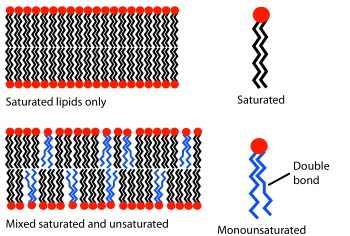
Ad una certa temperatura, un doppio strato lipidico può esistere sia in fase liquida che solida (gel). Tutti i lipidi posseggono una temperatura caratteristica a cui passano dallo stato di gel allo stato liquido (punto di fusione). In entrambe le fasi le molecole lipidiche sono ostacolate nel flip-flop, ma in fase liquida i singoli lipidi cambiano posizione con i loro vicini continuamente, milioni di volte al secondo. Questa scambio casuale permette ai lipidi di diffondere e in questo modo spostarsi sulla superficie della membrana.[17] Al contrario, i lipidi in fase di gel sono bloccati nella loro posizione.
Il comportamento di fase dei doppi strati lipidici è determinato in gran parte dall'intensità delle forze di interazione attrattive tra le molecole lipidiche adiacenti, in gran parte forze di dispersione di London e interazioni idrofobiche fra le catene idrocarburiche. Lipidi con code più lunghe dispongono di una maggiore superficie di interazione e di conseguenza interagiscono maggiormente tra di loro, riducendo la mobilità molecolare. Per questo, ad una data temperatura, un lipide con code corte risulterà più fluido di un lipide con code lunghe.[7] La temperatura di fusione può anche essere influenzata dal grado di insaturazione delle code lipidiche. Un doppio legame insaturo può produrre una piega nella catena alifatica, interrompendo l'impaccamento lipidico. Questa interruzione crea ulteriori spazi liberi all'interno del doppio strato che permettono una maggiore flessibilità alle catene adiacenti.[7] Un esempio di questo effetto si può notare nella vita di ogni giorno, dal momento che il burro, che ha un'elevata percentuale di grassi saturi, è solido a temperatura ambiente, mentre l'olio vegetale, che è principalmente insaturo, è liquido.
La maggior parte delle membrane naturali sono un complesso miscuglio di molecole lipidiche differenti. Se alcuni dei componenti del doppio strato sono liquidi ad una data temperatura, mentre altri sono in fase di gel, le due fasi possono coesistere in regioni spaziali separate, in maniera abbastanza simile ad un iceberg che galleggia nell'oceano. Questa separazione di fase ha un ruolo fondamentale nei fenomeni biochimici, perché componenti della membrana, come le proteine, possono separarsi in una o nell'altra fase.[18] e per questo motivo essere concentrato o attivate localmente. Un componente particolarmente importante di molti sistemi con fasi differenti è il colesterolo, che modula la permeabilità, la resistenza meccanica e le interazioni biochimiche del doppio strato.
Chimica di superficie[modifica | modifica wikitesto]
Mentre le code lipidiche modulano principalmente il comportamento di fase del doppio strato, sono i gruppi di testa che determinano la sua chimica di superficie. La maggior parte dei doppi strati naturali sono composti principalmente da fosfolipidi, sebbene anche gli sfingolipidi, come la sfingomielina, e gli steroli, come il colesterolo, siano componenti importanti. Dei fosfolipidi, il gruppo di testa più comune è la fosfatidilcolina, che rappresenta circa la metà dei fosfolipidi nella maggior parte delle cellule di mammifero.[19] Essa è un gruppo di testa zwitterionico, dal momento che presenta una carica negativa sul gruppo fosfato e una positiva su quello amminico, ma nessuna carica netta, in quanto queste cariche locali si bilanciano a vicenda.
Altri gruppi di testa sono anche presenti in diverse proporzioni e possono includere fosfatidilserina, fosfatidiletanolammina e fosfatidilglicerolo. Questi gruppi di testa alternativi spesso conferiscono alla membrana specifiche proprietà biologiche che sono altamente dipendenti dal contesto. Ad esempio, la presenza di fosfatidilserina sulla faccia extracellulare degli eritrociti può indurre apoptosi,[20] mentre nelle vescicole della cartilagine epifisaria è necessaria per la nucleazione dei cristalli di idrossiapatite e perciò alla mineralizzazione dell'osso.[21][22] Diversamente dalla fosfatidilcolina, alcuni degli altri gruppi di testa portano una carica netta, che può alterare le interazioni elettrostatiche di piccole molecole con il doppio strato.[23]
Funzioni biologiche[modifica | modifica wikitesto]
Contenimento e distinzione[modifica | modifica wikitesto]
Il ruolo principale del doppio strato lipidico in biologia è quello di separare compartimenti acquosi dall'ambiente esterno. Senza una qualche forma di barriera che delinei ciò che è proprio (self) da ciò che non lo è (non-self), è difficile persino definire il concetto di un organismo o della vita. Questa barriera assume la forma di un doppio strato lipidico in tutte le forme conosciute di vita, eccetto che in poche specie di archei che utilizzano un monostrato lipidico specialmente adattato.[4] È stata perfino avanzata l'ipotesi che le primissime forme di vita possano esser state delle semplici vescicole lipidiche, avendo ipoteticamente come unica capacità biosintetica quella di produrre fosfolipidi.[24] La capacità di separazione del doppio strato lipidico si basa sul fatto che le molecole idrofiliche non possono oltrepassare facilmente il suo nucleo idrofobico. (vedi oltre)
I procarioti posseggono un unico doppio strato lipidico, la membrana cellulare (conosciuta anche come membrana plasmatica). Molti procarioti posseggono anche una parete cellulare, ma essa è composta da proteine o da lunghe catene di carboidrati. Gli eucarioti, invece, posseggono numerosi organelli, tra cui il nucleo, i mitocondri, i lisosomi e il reticolo endoplasmatico. Tutti questi compartimenti subcellulari sono circondati da uno o più doppi strati lipidici e insieme assommano la maggioranza della superficie membranosa della cellula. Negli epatociti, ad esempio, la membrana plasmatica rappresenta solo il due percento della superficie totale di doppio strato, mentre il reticolo endoplasmatico ne rappresenta più della metà e i mitocondri un ulteriore trenta percento.[25]
Segnalazione[modifica | modifica wikitesto]
Probabilmente la forma più nota forma di segnalazione cellulare è la trasmissione sinaptica, per mezzo della quale un impulso nervoso che ha raggiunto l'estremità dell'assone di un neurone è trasferito a un neurone adiacente attraverso il rilascio di vescicole sinaptiche piene di neurotrasmettitori. Queste vescicole si fondono con la membrana cellulare al terminale pre-sinaptico e rilasciano i loro contenuti all'esterno della cellula, nello spazio sinaptico. Essi diffondono e raggiungono il terminale post-sinaptico.
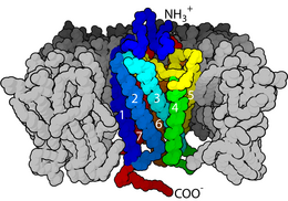
I doppi strati lipidici sono anche coinvolti nella trasduzione del segnale in quanto ospitano le proteine integrali di membrana. Questa è una classe estremamente vasta ed importante di biomolecole. È stato stimato che fino ad un terzo del proteoma umano possa essere costituito da proteine di membrana.[26] Alcune di queste proteine sono legate all'esterno della membrana cellulare. Un esempio di queste è la proteina CD59, che identifica le cellule come self (ossia appartenenti all'organismo) e perciò inibisce la loro distruzione da parte del sistema immunitario. Il virus HIV elude il sistema immunitario in parte innestando tali proteine sulla sua superficie, prelevandole dalla membrana ospite.[25] Alcune proteine, invece, attraversano completamente il doppio strato e servono a tramettere singoli eventi di segnalazione dall'esterno all'interno della cellula. La classe più comune di questo tipo di proteine è quella dei recettori accoppiati a proteine G (GPCR). Essi sono responsabili per la maggior parte della capacità della cellula di essere sensibile all'ambiente vicino e, a causa di questo ruolo importante, circa il 40% dei moderni farmaci sono indirizzati verso di essi.[27]
Oltre ai processi mediati da proteine e da soluzioni, i doppi strati lipidici possono partecipare direttamente alla segnalazione. Un esempio classico di questa capacità è dato dalla fagocitosi attivata da fosfatidilserina. Normalmente, la fosfatidilserina è distribuita asimmetricamente nella membrana plasmatica, ed è presente solo nel versante citosolico. Durante la morte cellulare programmata (apoptosi) un enzima chiamato scramblasi riequilibra questa distribuzione, mostrando il fosfolipide sul versante extracellulare. La sua presenza innaturale in esso avvia la fagocitosi, che rimuove la cellula ormai morta o morente.
Metodi di caratterizzazione[modifica | modifica wikitesto]

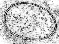
Il doppio strato lipidico è una struttura molto difficile da studiare, a causa della suo ridotto spessore e della sua fragilità. Nonostante questi problemi, sono state sviluppate molte tecniche negli ultimi settant'anni che permettono lo studio della sua struttura e delle sue funzioni.
Misure elettriche sono un modo semplice per definire una funzione importante del doppio strato: la sua capacità di separare ed impedire il flusso di ioni in soluzione. Applicando una differenza di potenziale attraverso la membrana e misurando la corrente elettrica che ne risulta, si può determinare la resistenza elettrica del doppio strato. Questa è solitamente abbastanza alta, dal momento che il nucleo idrofobico è impermeabile alle specie cariche. La presenza di pochi buchi di grandezza nanometrica porta ad un notevole aumento della corrente che attraversa la membrana.[28] La sensibilità di questo sistema è tale che può essere definita persino l'attività di un singolo canale ionico.[29]
Le misurazioni elettriche, però, non forniscono un'immagine "reale" della membrana. Per ottenerla, non sono sufficienti i microscopi tradizionali, ma sono spesso utilizzati microscopi a fluorescenza. Un campione viene eccitato con luce di una particolare lunghezza d'onda ed è osservato in una differente lunghezza d'onda, in modo tale che solo le molecole fluorescenti con un profilo di eccitazione ed emissione che corrisponde possano essere osservate. I doppi strati lipidici naturali non sono fluorescenti, viene quindi utilizzato un colorante che si lega selettivamente alle molecole a cui si è interessati. La risoluzione è solitamente limitata a poche centinaia di nanometri, molto più piccola di una tipica cellula, ma molto più grande dello spessore di un doppio strato lipidico.


La microscopia elettronica offre una maggiore risoluzione di immagine. In un microscopio elettronico un fascio focalizzato di elettroni interagisce con il campione. Insieme a tecniche di rapido congelamento, la microscopia elettronica è stata utilizzata anche per studiare i meccanismi di trasporto intracellulare ed intercellulare, per esempio per dimostrare che le vescicole esocitiche sono il mezzo di rilascio chimico nelle sinapsi.[31]
Un nuovo metodo di studio dei doppi strati lipidici è la microscopia a forza atomica (AFM). Invece di utilizzare un raggio di luce o di particelle, una piccolissima punta scansiona la superficie mediante un contatto fisico diretto con il doppio strato e muovendosi su di esso, come una puntina di giradischi. AFM is a promising technique because it has the potential to image with nanometer resolution at room temperature and even under water or physiological buffer, conditions necessary for natural bilayer behavior. Utilizing this capability, AFM has been used to examine dynamic bilayer behavior including the formation of transmembrane pores (holes)[30] and phase transitions in supported bilayers.[32] Another advantage is that AFM doesn't require fluorescent or isotopic labeling of the lipids, since the probe tip interacts mechanically with the bilayer surface. Because of this, the same scan can image both lipids and associated proteins, sometimes even with single-molecule resolution.[30][33] AFM can also probe the mechanical nature of lipid bilayers.[34]
Lipid bilayers exhibit high levels of birefringence where the refractive index in the plane of the bilayer differs from that perpendicular by as much as 0.1 refractive index units. This has been used to characterise the degree of order and disruption in bilayers using dual polarisation interferometry to understand mechanisms of protein interaction.
Transport across the bilayer[modifica | modifica wikitesto]
Passive diffusion[modifica | modifica wikitesto]
Most polar molecules have low solubility in the hydrocarbon core of a lipid bilayer and consequently have low permeability coefficients across the bilayer. This effect is particularly pronounced for charged species, which have even lower permeability coefficients than neutral polar molecules.[35] Anions typically have a higher rate of diffusion through bilayers than cations.[36][37] Compared to ions, water molecules actually have a relatively large permeability through the bilayer, as evidenced by osmotic swelling. When a cell or vesicle with a high interior salt concentration is placed in a solution with a low salt concentration it will swell and eventually burst. Such a result would not be observed unless water was able to pass through the bilayer with relative ease. The anomalously large permeability of water through bilayers is still not completely understood and continues to be the subject of active debate.[38] Uncharged apolar molecules diffuse through lipid bilayers many orders of magnitude faster than ions or water. This applies both to fats and organic solvents like chloroform and ether. Regardless of their polar character larger molecules diffuse more slowly across lipid bilayers than small molecules.[39]
Ion pumps and channels[modifica | modifica wikitesto]
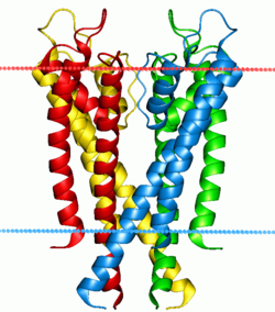
Two special classes of protein deal with the ionic gradients found across cellular and sub-cellular membranes in nature- ion channels and ion pumps. Both pumps and channels are integral membrane proteins that pass through the bilayer, but their roles are quite different. Ion pumps are the proteins that build and maintain the chemical gradients by utilizing an external energy source to move ions against the concentration gradient to an area of higher chemical potential. The energy source can be ATP, as is the case for the Na+-K+ ATPase. Alternatively, the energy source can be another chemical gradient already in place, as in the Ca2+/Na+ antiporter. It is through the action of ion pumps that cells are able to regulate pH via the pumping of protons.
In contrast to ion pumps, ion channels do not build chemical gradients but rather dissipate them in order to perform work or send a signal. Probably the most familiar and best studied example is the voltage-gated Na+ channel, which allows conduction of an action potential along neurons. All ion pumps have some sort of trigger or “gating” mechanism. In the previous example it was electrical bias, but other channels can be activated by binding a molecular agonist or through a conformational change in another nearby protein.[40]

Endocytosis and exocytosis[modifica | modifica wikitesto]
Template:Also Some molecules or particles are too large or too hydrophilic to effectively pass through a lipid bilayer. Other molecules could pass through the bilayer but must be transported rapidly in such large numbers that channel-type transport is impractical. In both cases these types of cargo can be moved across the cell membrane through fusion or budding of vesicles. When a vesicle is produced inside the cell and fuses with the plasma membrane to release its contents into the extracellular space this process is known as exocytosis. In the reverse process a region of the cell membrane will dimple inwards and eventually pinch off, enclosing a portion of the extracellular fluid to transport it into the cell. Endocytosis and exocytosis rely on very different molecular machinery to function, but the two processes are intimately linked and could not work without each other. The primary mechanism this interdependence is the sheer volume of lipid material involved.[41] In a typical cell, an area of bilayer equivalent to the entire plasma membrane will travel through the endocytosis/exocytosis cycle in about half an hour.[42] If these two processes were not balancing each other the cell would either balloon outward to an unmanageable size or completely deplete its plasma membrane within a matter of minutes.
Electroporation[modifica | modifica wikitesto]
Electroporation is the rapid increase in bilayer permeability induced by the application of a large artificial electric field across the membrane. Experimentally, electroporation is used to introduce hydrophilic molecules into cells. It is a particularly useful technique for large highly charged molecules such as DNA which would never passively diffuse across the hydrophobic bilayer core.[43] Because of this, electroporation is one of the key methods of transfection as well as bacterial transformation. It has even been proposed that electroporation resulting from lightning strikes could be a mechanism of natural horizontal gene transfer.[44]
This increase in permeability primarily affects transport of ions and other hydrated species, indicating that the mechanism is the creation of nm-scale water-filled holes in the membrane. Although electroporation and dielectric breakdown both result from application of an electric field, the mechanisms involved are fundamentally different. In dielectric breakdown the barrier material is ionized, creating a conductive pathway. The material alteration is thus chemical in nature. In contrast, during electroporation the lipid molecules are not chemically altered but simply shift position, opening up a pore which acts as the conductive pathway through the bilayer as it is filled with water.
Mechanics[modifica | modifica wikitesto]

Lipid bilayers are large enough structures to have some of the mechanical properties of liquids or solids. The area compression modulus Ka, bending modulus Kb, and edge energy , can be used to describe them. Solid lipid bilayers also have a shear modulus, but like any liquid, the shear modulus is zero for fluid bilayers. These mechanical properties affect how the membrane functions. Ka and Kb affect the ability of proteins and small molecules to insert into the bilayer,[45][46] and bilayer mechanical properties have been shown to alter the function of mechanically activated ion channels.[47] Bilayer mechanical properties also govern what types of stress a cell can withstand without tearing. Although lipid bilayers can easily bend, most cannot stretch more than a few percent before rupturing.[48]
As discussed in the Structure and organization section, the hydrophobic repulsion between lipid tails and water is the primary force holding lipid bilayers together. Thus, the elastic modulus of the bilayer is primarily determined by how much extra area is exposed to water when the lipid molecules are stretched apart.[49] It is not surprising given this understanding of the forces involved that studies have shown that Ka varies strongly with solution conditions[50] but only weakly with tail length and unsaturation.[7] Because the forces involved are so small, it is difficult to experimentally determine Ka. Most techniques require sophisticated microscopy and very sensitive measurement equipment.[34][51]
In contrast to Ka, which is a measure of how much energy is needed to stretch the bilayer, Kb is a measure of how much energy is needed to bend or flex the bilayer. Formally, bending modulus is defined as the energy required to deform a membrane from its intrinsic curvature to some other curvature. Intrinsic curvature is defined by the ratio of the diameter of the head group to that of the tail group. For two-tailed PC lipids, this ratio is nearly one so the intrinsic curvature is nearly zero. If a particular lipid has too large a deviation from zero intrinsic curvature it will not form a bilayer and will instead form other phases such as micelles or inverted micelles. Typically, Kb is not measured experimentally but rather is calculated from measurements of Ka and bilayer thickness, since the three parameters are related.
is a measure of how much energy it takes to expose a bilayer edge to water by tearing the bilayer or creating a hole in it. The origin of this energy is the fact that creating such an interface exposes some of the lipid tails to water, but the exact orientation of these border lipids is unknown. There is some evidence that both hydrophobic (tails straight) and hydrophilic (heads curved around) pores can coexist.[52]
Fusion[modifica | modifica wikitesto]

Fusion is the process by which two lipid bilayers merge, resulting in one connected structure. If this fusion proceeds completely through both leaflets of both bilayers, a water-filled bridge is formed and the solutions contained by the bilayers can mix. Alternatively, if only one leaflet from each bilayer is involved in the fusion process, the bilayers are said to be hemifused. Fusion is involved in many cellular processes, particularly in eukaryotes since the eukaryotic cell is extensively sub-divided by lipid bilayer membranes. Exocytosis, fertilization of an egg by sperm and transport of waste products to the lysozome are a few of the many eukaryotic processes that rely on some form of fusion. Even the entry of pathogens can be governed by fusion, as many bilayer-coated viruses have dedicated fusion proteins to gain entry into the host cell.
There are four fundamental steps in the fusion process.[19] First, the involved membranes must aggregate, approaching each other to within several nanometers. Second, the two bilayers must come into very close contact (within a few angstroms). To achieve this close contact, the two surfaces must become at least partially dehydrated, as the bound surface water normally present causes bilayers to strongly repel. The presence of ions, particularly divalent cations like magnesium and calcium, strongly affects this step.[53][54] One of the critical roles of calcium in the body is regulating membrane fusion. Third, a destabilization must form at one point between the two bilayers, locally distorting their structures. The exact nature of this distortion is not known. One theory is that a highly curved "stalk" must form between the two bilayers.[55] Proponents of this theory believe that it explains why phosphatidylethanolamine, a highly curved lipid, promotes fusion.[56] Finally, in the last step of fusion, this point defect grows and the components of the two bilayers mix and diffuse away from the site of contact.


The situation is further complicated when considering fusion in vivo since biological fusion is almost always regulated by the action of membrane-associated proteins. The first of these proteins to be studied were the viral fusion proteins, which allow an enveloped virus to insert its genetic material into the host cell (enveloped viruses are those surrounded by a lipid bilayer; some others have only a protein coat).Eukaryotic cells also use fusion proteins, the best studied of which are the SNAREs. SNARE proteins are used to direct all vesicular intracellular trafficking. Despite years of study, much is still unknown about the function of this protein class. In fact, there is still an active debate regarding whether SNAREs are linked to early docking or participate later in the fusion process by facilitating hemifusion.[57]
In studies of molecular and cellular biology it is often desirable to artificially induce fusion. The addition of polyethylene glycol (PEG) causes fusion without significant aggregation or biochemical disruption. This procedure is now used extensively, for example by fusing B-cells with melanoma cells.[58] The resulting “hybridoma” from this combination expresses a desired antibody as determined by the B-cell involved, but is immortalized due to the melanoma component. Fusion can also be artificially induced through electroporation in a process known as electrofusion. It is believed that this phenomenon results from the energetically active edges formed during electroporation, which can act as the local defect point to nucleate stalk growth between two bilayers.[59]
Model systems[modifica | modifica wikitesto]
Lipid bilayers can be created artificially in the lab to allow researchers to perform experiments that cannot be done with natural bilayers. These synthetic systems are called model lipid bilayers. There are many different types of model bilayers, each having experimental advantages and disadvantages. They can be made with either synthetic or natural lipids. Some of the most common systems are described below.

Black lipid membranes (BLM)[modifica | modifica wikitesto]
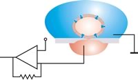
The earliest model bilayer system developed was the “painted” bilayer, also known as a “black lipid membrane.” The term “painted” refers to the process by which these bilayers are made. The term “black” bilayer refers to the fact that they are dark in reflected light because the thickness of the membrane is only a few nanometers, so light reflecting off the back face destructively interferes with light reflecting off the front face. To make a BLM, a small aperture is created in a hydrophobic material such as Teflon. A solution of lipids dissolved in an organic solvent is then applied with a brush or a syringe across the aperture.[60] Black lipid membranes are well suited to electrical measurements like resistance (typically gigaOhms or higher for intact bilayers) and capacitance (~2µF/cm2). Electrical characterization has been particularly important in the study of voltage gated ion channels, which can be inserted into a BLM by coating them with a detergent and mixing them into the solution surrounding the BLM.
Supported lipid bilayers (SLB)[modifica | modifica wikitesto]

A supported bilayer is a sheet that lays flat on a solid substrate such that only the upper face of the bilayer is exposed to free solution. One advantage of this layout is its stability. SLBs will remain largely intact even when subject to high flow rates or vibration and, unlike black lipid membranes, the presence of holes will not destroy the entire bilayer. Because of this stability, experiments lasting weeks and even months are possible with supported bilayers while BLM experiments are usually limited to hours.[61]

Another advantage of the supported bilayer is that, because it is on a flat hard surface, it is amenable to a number of characterization tools which would be impossible or would offer lower resolution if performed on a freely floating sample. One of the clearest examples of this advantage is the use of mechanical probing techniques which require a direct physical interaction with the sample such as Atomic force microscopy (AFM). Many modern fluorescence microscopy techniques such as total internal reflection fluorescence microscopy (TIRF) and surface plasmon resonance (SPR) also require a rigidly-supported planar surface.

One of the primary limitations of supported bilayers is the possibility of unwanted interactions with the substrate. Although supported bilayers generally do not directly touch the substrate surface, they are separated by only a very thin water gap. The size and nature of this gap depends on the substrate material[62] and lipid species but is generally about 1 nm.[63][64] Because this layer is so thin, there are often problem when incorporating integral membrane proteins, which can become denatured on the substrate surface.[65] One approach to circumvent this problem is to support the bilayer on a loose network of hydrated polymers or a hydrogel which acts as a spacer between lipids and substrate.[66]
Vesicles[modifica | modifica wikitesto]
A vesicle is a lipid bilayer rolled up into a spherical shell, enclosing a small amount of water and separating it from the water outside the vesicle. Vesicles are relatively easy to make; if a sample of dehydrated lipid is exposed to water it will spontaneously form vesicles.[67] Since artificial vesicles can be made in large quantities they are suitable for bulk material studies such as x-ray diffraction to determine lattice spacing[68] and differential scanning calorimetry to determine phase transitions.[8] In spite of this fluorescent labeling it is often difficult to perform detailed imaging on SUVs simply because they are so small. To combat this problem researchers have developed the giant unilamellar vesicle (GUV). GUVs are large enough (several tens of micrometres) to study with traditional florescence microscopy. Compared to supported bilayers, GUVs present a more “natural” environment since there is no nearby solid surface to induce defects or denature proteins. However, GUVs are relatively fragile, time consuming to make and can only be produced in limited yield compared to SUVs.
Commercial applications[modifica | modifica wikitesto]

To date, the most successful commercial application of lipid bilayers has been the use of liposomes for drug delivery, especially for cancer treatment. (Note- the term “liposome” is essentially synonymous with “vesicle” except that vesicle is a general term for the structure whereas liposome only refers to artificial, not natural vesicles) The basic idea of liposomal drug delivery is that the drug is encapsulated in solution inside the liposome then injected into the patient. These drug-loaded liposomes travel through the system until they bind at the target site and rupture, releasing the drug. In theory, liposomes should make an ideal drug delivery system since they can isolate nearly any hydrophilic drug, can be grafted with molecules to target specific tissues and can be relatively non-toxic since the body possesses biochemical pathways for degrading lipids.[69]
The first generation of drug delivery liposomes had a simple lipid composition and suffered from several limitations. Circulation in the bloodstream was extremely limited due to both renal clearing and phagocytosis. Refinement of the lipid composition to tune fluidity, surface charge density and surface hydration resulted in vesicles that adsorb fewer proteins from serum and thus are less readily recognized by the immune system.[70] The most significant advance in this area was the grafting of polyethylene glycol (PEG) onto the liposome surface to produce “stealth” vesicles which circulate over long times without immune or renal clearing.[71]
The first stealth liposomes were passively targeted at tumor tissues. Because tumors induce rapid and uncontrolled angiogenesis they are especially “leaky” and allow liposomes to exit the bloodstream at a much higher rate than normal tissue would.[72] More recently, work has been undertaken to graft antibodies or other molecular markers onto the liposome surface in the hope of actively binding them to a specific cell or tissue type.[73] Some examples of this approach are already in clinical trials.[74]
Another potential application of lipid bilayers is the field of biosensors. Since the lipid bilayer is the barrier between the interior and exterior of the cell it is also the site of extensive signal transduction. Researchers over the years have tried to harness this potential to develop a bilayer-based device for clinical diagnosis or bioterrorism detection. Progress has been slow in this area and, although a few companies have developed automated lipid-based detection systems, they are still targeted at the research community. These include Biacore Life Sciences, which offers a disposable chip for utilizing lipid bilayers in studies of binding kinetics[75] and Nanion Inc which has developed an automated patch clamping system.[76] Other, more exotic applications are also being pursued such as the use of lipid bilayer membrane pores for DNA sequencing by Oxford Nanolabs. To date, this technology has not proven commercially viable.
History[modifica | modifica wikitesto]
By the early twentieth century scientists had come to believe that cells are surrounded by a thin oil-like barrier,[77] but the structural nature of this membrane was not known. Two experiments in 1925 laid the groundwork to fill in this gap. By measuring the capacitance of erythrocyte solutions, Hugo Fricke determined that the cell membrane was 3.3 nm thick.[78] Although the results of this experiment were accurate, Fricke misinterpreted the data to mean that the cell membrane is a single molecular layer. Prof. Dr. Evert Gorter[79] (1881-1954) and F. Grendel of Leiden University approached the problem from a different perspective, spreading the erythrocyte lipids as a monolayer on a Langmuir-Blodgett trough. When they compared the area of the monolayer to the surface area of the cells, they found a ratio of two to one.[80] Later analyses showed several errors and incorrect assumptions with this experiment but, serendipitously, these errors canceled out and from this flawed data Gorter and Grendel drew the correct conclusion- that the cell membrane is a lipid bilayer.[19]
This theory was confirmed through the use of electron microscopy in the late 1950s. Although he did not publish the first electron microscopy study of lipid bilayers[81] J. David Robertson was the first to assert that the two dark electron-dense bands were the headgroups and associated proteins of two apposed lipid monolayers.[82][83] In this body of work, Robertson put forward the concept of the “unit membrane.” This was the first time the bilayer structure had been universally assigned to all cell membranes as well as organelle membranes.
Around the same time the development of model membranes confirmed that the lipid bilayer is a stable structure that can exist independently of proteins. By “painting” a solution of lipid in organic solvent across an aperture, Mueller and Rudin were able to create an artificial bilayer and determine that this exhibited lateral fluidity, high electrical resistance and self-healing in response to puncture,[60] all of which are properties of a natural cell membrane. A few years later, Alec Bangham showed that bilayers, in the form of lipid vesicles, could also be formed simply by exposing a dried lipid sample to water.[67] This was an important advance since it demonstrated that lipid bilayers form spontaneously via self assembly and do not require a patterned support structure.
See also[modifica | modifica wikitesto]
References[modifica | modifica wikitesto]
- ^ a b Lewis BA, Engelman DM, Lipid bilayer thickness varies linearly with acyl chain length in fluid phosphatidylcholine vesicles, in J. Mol. Biol., vol. 166, n. 2, maggio 1983, pp. 211–7, DOI:10.1016/S0022-2836(83)80007-2, PMID 6854644.
- ^ Zaccai G, Blasie JK, Schoenborn BP, Neutron Diffraction Studies on the Location of Water in Lecithin Bilayer Model Membranes (PDF), in Proc. Natl. Acad. Sci. U.S.A., vol. 72, n. 1, gennaio 1975, pp. 376–380, DOI:10.1073/pnas.72.1.376, PMID 16592215.
- ^ Nagle JF, Tristram-Nagle S, Structure of lipid bilayers (PDF), in Biochim. Biophys. Acta, vol. 1469, n. 3, novembre 2000, pp. 159–95, DOI:10.1016/S0304-4157(00)00016-2, PMID 11063882.
- ^ a b Parker J, Madigan MT, Brock TD, Martinko JM, Brock biology of microorganisms, 10ª ed., Englewood Cliffs, N.J, Prentice Hall, 2003, ISBN 0-13-049147-0.
- ^ Marsh D, Polarity and permeation profiles in lipid membranes (PDF), in Proc. Natl. Acad. Sci. U.S.A., vol. 98, n. 14, luglio 2001, pp. 7777–82, DOI:10.1073/pnas.131023798, PMID 11438731.
- ^ Marsh D, Membrane water-penetration profiles from spin labels, in Eur. Biophys. J., vol. 31, n. 7, dicembre 2002, pp. 559–62, DOI:10.1007/s00249-002-0245-z, PMID 12602343.
- ^ a b c d Rawicz W, Olbrich KC, McIntosh T, Needham D, Evans E, Effect of chain length and unsaturation on elasticity of lipid bilayers (PDF), in Biophys. J., vol. 79, n. 1, luglio 2000, pp. 328–39, DOI:10.1016/S0006-3495(00)76295-3, PMID 10866959.
- ^ a b Trauble H, Haynes DH, The volume change in lipid bilayer lamellae at the crystalline-liquid crystalline phase transition, in Chem. Phys. Lipids, vol. 7, n. 4, dicembre 1971, pp. 324–35, DOI:10.1016/0009-3084(71)90010-7. Errore nelle note: Tag
<ref>non valido; il nome "Trauble1971" è stato definito più volte con contenuti diversi - ^ Verkleij AJ, Zwaal RF, Roelofsen B, Comfurius P, Kastelijn D, van Deenen LL, The asymmetric distribution of phospholipids in the human red cell membrane. A combined study using phospholipases and freeze-etch electron microscopy, in Biochim. Biophys. Acta, vol. 323, n. 2, ottobre 1973, pp. 178–93, DOI:10.1016/0005-2736(73)90143-0, PMID 4356540.
- ^ Bell RM, Ballas LM, Coleman RA, Lipid topogenesis (PDF), in J. Lipid Res., vol. 22, n. 3, 1° marzo 1981, pp. 391–403, PMID 7017050.
- ^ Rothman JE, Kennedy EP, Rapid transmembrane movement of newly synthesized phospholipids during membrane assembly (PDF), in Proc. Natl. Acad. Sci. U.S.A., vol. 74, n. 5, maggio 1977, pp. 1821–5, DOI:10.1073/pnas.74.5.1821, PMID 405668.
- ^ Kornberg RD, McConnell HM, Inside-outside transitions of phospholipids in vesicle membranes, in Biochemistry, vol. 10, n. 7, marzo 1971, pp. 1111–20, DOI:10.1021/bi00783a003, PMID 4324203.
- ^ Litman BJ, Determination of molecular asymmetry in the phosphatidylethanolamine surface distribution in mixed phospholipid vesicles, in Biochemistry, vol. 13, n. 14, luglio 1974, pp. 2844–8, DOI:10.1021/bi00711a010, PMID 4407872.
- ^ Crane JM, Kiessling V, Tamm LK, Measuring lipid asymmetry in planar supported bilayers by fluorescence interference contrast microscopy, in Langmuir, vol. 21, n. 4, febbraio 2005, pp. 1377–88, DOI:10.1021/la047654w, PMID 15697284.
- ^ Kalb E, Frey S, Tamm LK, Formation of supported planar bilayers by fusion of vesicles to supported phospholipid monolayers, in Biochim. Biophys. Acta, vol. 1103, n. 2, gennaio 1992, pp. 307–16, DOI:10.1016/0005-2736(92)90101-Q, PMID 1311950.
- ^ Lin WC, Blanchette CD, Ratto TV, Longo ML, Lipid asymmetry in DLPC/DSPC-supported lipid bilayers: a combined AFM and fluorescence microscopy study (PDF), in Biophys. J., vol. 90, n. 1, gennaio 2006, pp. 228–37, DOI:10.1529/biophysj.105.067066, PMID 16214871.
- ^ Berg, Howard C, Random walks in biology, Extended Paperback, Princeton, N.J, Princeton University Press, 1993, ISBN 0-691-
ISBNnon valido (aiuto), .... - ^ Dietrich C, Volovyk ZN, Levi M, Thompson NL, Jacobson K, Partitioning of Thy-1, GM1, and cross-linked phospholipid analogs into lipid rafts reconstituted in supported model membrane monolayers (PDF), in Proc. Natl. Acad. Sci. U.S.A., vol. 98, n. 19, settembre 2001, pp. 10642–7, DOI:10.1073/pnas.191168698, PMID 11535814.
- ^ a b c Yeagle, Philip, The membranes of cells, 2nd, Boston, Academic Press, 1993, ISBN 0-12-769041-7.
- ^ Fadok VA, Bratton DL, Frasch SC, Warner ML, Henson PM, The role of phosphatidylserine in recognition of apoptotic cells by phagocytes (PDF), in Cell Death Differ, vol. 5, n. 7, luglio 1998, pp. 551–62, DOI:10.1038/sj.cdd.4400404, PMID 10200509.
- ^ Anderson HC, Garimella R, Tague SE, The role of matrix vesicles in growth plate development and biomineralization, in Front. Biosci., vol. 10, gennaio 2005, pp. 822–37, DOI:10.2741/1576, PMID 15569622.
- ^ Eanes ED, Hailer AW, Calcium phosphate precipitation in aqueous suspensions of phosphatidylserine-containing anionic liposomes, in Calcif. Tissue Int., vol. 40, n. 1, gennaio 1987, pp. 43–8, DOI:10.1007/BF02555727, PMID 3103899.
- ^ Kim J, Mosior M, Chung LA, Wu H, McLaughlin S, Binding of peptides with basic residues to membranes containing acidic phospholipids (PDF), in Biophys. J., vol. 60, n. 1, luglio 1991, pp. 135–48, DOI:10.1016/S0006-3495(91)82037-9, PMID 1883932.
- ^ Koch AL, Primeval cells: possible energy-generating and cell-division mechanisms, in J. Mol. Evol., vol. 21, n. 3, aprile 1984, pp. 270–7, DOI:10.1007/BF02102359, PMID 6242168.
- ^ a b Alberts, Bruce, Molecular biology of the cell, 4ª ed., New York, Garland Science, 2002, ISBN 0-8153-4072-9.
- ^ Martelli PL, Fariselli P, Casadio R, An ENSEMBLE machine learning approach for the prediction of all-alpha membrane proteins (PDF), in Bioinformatics, vol. 19, Suppl 1, 2003, pp. i205–11, DOI:10.1093/bioinformatics/btg1027, PMID 12855459.
- ^ Filmore D, It's A GPCR World (PDF), in Modern Drug Discovery, vol. 7, n. 11, novembre 2004, pp. 24–9.
- ^ Melikov KC, Frolov VA, Shcherbakov A, Samsonov AV, Chizmadzhev YA, Chernomordik LV, Voltage-induced nonconductive pre-pores and metastable single pores in unmodified planar lipid bilayer (PDF), in Biophys. J., vol. 80, n. 4, aprile 2001, pp. 1829–36, DOI:10.1016/S0006-3495(01)76153-X, PMID 11259296.
- ^ Neher E, Sakmann B, Single-channel currents recorded from membrane of denervated frog muscle fibres, in Nature, vol. 260, n. 5554, aprile 1976, pp. 799–802, DOI:10.1038/260799a0, PMID 1083489.
- ^ a b c Y. Roiter, M. Ornatska, A. R. Rammohan, J. Balakrishnan, D. R. Heine, and S. Minko, Interaction of Nanoparticles with Lipid Membrane, Nano Letters, vol. 8, iss. 3, pp. 941–944 (2008).
- ^ Heuser JE, Reese TS, Dennis MJ, Jan Y, Jan L, Evans L, Synaptic vesicle exocytosis captured by quick freezing and correlated with quantal transmitter release (PDF), in J. Cell Biol., vol. 81, n. 2, maggio 1979, pp. 275–300, DOI:10.1083/jcb.81.2.275, PMID 38256.
- ^ Tokumasu F, Jin AJ, Dvorak JA, Lipid membrane phase behavior elucidated in real time by controlled environment atomic force microscopy, in J. Electron Micros., vol. 51, n. 1, 2002, pp. 1–9.
- ^ Richter RP, Brisson A, Characterization of lipid bilayers and protein assemblies supported on rough surfaces by atomic force microscopy, in Langmuir, vol. 19, 2003, pp. 1632–40, DOI:10.1021/la026427w.
- ^ a b Steltenkamp S, Müller MM, Deserno M, Hennesthal C, Steinem C, Janshoff A, Mechanical properties of pore-spanning lipid bilayers probed by atomic force microscopy, in Biophys. J., vol. 91, n. 1, July 2006, pp. 217–26, DOI:10.1529/biophysj.106.081398, PMC 1479081, PMID 16617084.
- ^ Chakrabarti AC, Permeability of membranes to amino acids and modified amino acids: mechanisms involved in translocation, in Amino Acids, vol. 6, 1994, pp. 213–29, DOI:10.1007/BF00813743, PMID 11543596.
- ^ Hauser H, Phillips MC, Stubbs M, Ion permeability of phospholipid bilayers, in Nature, vol. 239, n. 5371, October 1972, pp. 342–4, DOI:10.1038/239342a0, PMID 12635233.
- ^ Papahadjopoulos D, Watkins JC, Phospholipid model membranes. II. Permeability properties of hydrated liquid crystals, in Biochim. Biophys. Acta, vol. 135, n. 4, September 1967, pp. 639–52, DOI:10.1016/0005-2736(67)90095-8, PMID 6048247.
- ^ Paula S, Volkov AG, Van Hoek AN, Haines TH, Deamer DW, Permeation of protons, potassium ions, and small polar molecules through phospholipid bilayers as a function of membrane thickness, in Biophys. J., vol. 70, n. 1, January 1996, pp. 339–48, DOI:10.1016/S0006-3495(96)79575-9, PMC 1224932, PMID 8770210.
- ^ Xiang TX, Anderson BD, The relationship between permeant size and permeability in lipid bilayer membranes, in J. Membr. Biol., vol. 140, n. 2, June 1994, pp. 111–22, PMID 7932645.
- ^ Gouaux E, Mackinnon R, Principles of selective ion transport in channels and pumps, in Science, vol. 310, n. 5753, December 2005, pp. 1461–5, DOI:10.1126/science.1113666, PMID 16322449.
- ^ Gundelfinger ED, Kessels MM, Qualmann B, Temporal and spatial coordination of exocytosis and endocytosis, in Nat. Rev. Mol. Cell Biol., vol. 4, n. 2, February 2003, pp. 127–39, DOI:10.1038/nrm1016, PMID 12563290.
- ^ Steinman RM, Brodie SE, Cohn ZA, Membrane flow during pinocytosis. A stereologic analysis, in J. Cell Biol., vol. 68, n. 3, March 1976, pp. 665–87, DOI:10.1083/jcb.68.3.665, PMC 2109655, PMID 1030706.
- ^ Neumann E, Schaefer-Ridder M, Wang Y, Hofschneider PH, Gene transfer into mouse lyoma cells by electroporation in high electric fields, in Embo J., vol. 1, n. 7, 1982, pp. 841–5, PMC 553119, PMID 6329708.
- ^ Demanèche S, Bertolla F, Buret F, et al., Laboratory-scale evidence for lightning-mediated gene transfer in soil, in Appl. Environ. Microbiol., vol. 67, n. 8, August 2001, pp. 3440–4, DOI:10.1128/AEM.67.8.3440-3444.2001, PMC 93040, PMID 11472916.
- ^ Garcia ML, Ion channels: gate expectations, in Nature, vol. 430, n. 6996, July 2004, pp. 153–5, DOI:10.1038/430153a, PMID 15241399.
- ^ McIntosh TJ, Simon SA, Roles of Bilayer Material Properties in Function and Distribution of Membrane Proteins, in Annu. Rev. Biophys. Biomol. Struct., vol. 35, 2006, pp. 177–98, DOI:10.1146/annurev.biophys.35.040405.102022.
- ^ Suchyna TM, Tape SE, Koeppe RE, Andersen OS, Sachs F, Gottlieb PA, Bilayer-dependent inhibition of mechanosensitive channels by neuroactive peptide enantiomers, in Nature, vol. 430, n. 6996, July 2004, pp. 235–40, DOI:10.1038/nature02743, PMID 15241420.
- ^ Hallett FR, Marsh J, Nickel BG, Wood JM, Mechanical properties of vesicles. II. A model for osmotic swelling and lysis, in Biophys. J., vol. 64, n. 2, February 1993, pp. 435–42, DOI:10.1016/S0006-3495(93)81384-5, PMC 1262346, PMID 8457669.
- ^ Boal, David H., Mechanics of the cell, Cambridge, UK, Cambridge University Press, 2001, ISBN 0-521-79681-4.
- ^ Rutkowski CA, Williams LM, Haines TH, Cummins HZ, The elasticity of synthetic phospholipid vesicles obtained by photon correlation spectroscopy, in Biochemistry, vol. 30, n. 23, June 1991, pp. 5688–96, DOI:10.1021/bi00237a008, PMID 2043611.
- ^ Evans E, Heinrich V, Ludwig F, Rawicz W, Dynamic tension spectroscopy and strength of biomembranes, in Biophys. J., vol. 85, n. 4, October 2003, pp. 2342–50, DOI:10.1016/S0006-3495(03)74658-X, PMC 1303459, PMID 14507698.
- ^ Weaver JC, Chizmadzhev YA, Theory of electroporation: A review, in Biochemistry and Bioenergetics, vol. 41, 1996, pp. 135–60, DOI:10.1016/S0302-4598(96)05062-3.
- ^ Papahadjopoulos D, Nir S, Düzgünes N, Molecular mechanisms of calcium-induced membrane fusion, in J. Bioenerg. Biomembr., vol. 22, n. 2, April 1990, pp. 157–79, DOI:10.1007/BF00762944, PMID 2139437.
- ^ Leventis R, Gagné J, Fuller N, Rand RP, Silvius JR, Divalent cation induced fusion and lipid lateral segregation in phosphatidylcholine-phosphatidic acid vesicles, in Biochemistry, vol. 25, n. 22, November 1986, pp. 6978–87, DOI:10.1021/bi00370a600, PMID 3801406.
- ^ Markin VS, Kozlov MM, Borovjagin VL, On the theory of membrane fusion. The stalk mechanism, in Gen. Physiol. Biophys., vol. 3, n. 5, October 1984, pp. 361–77, PMID 6510702.
- ^ Chernomordik LV, Kozlov MM, Protein-lipid interplay in fusion and fission of biological membranes, in Annu. Rev. Biochem., vol. 72, 2003, pp. 175–207, DOI:10.1146/annurev.biochem.72.121801.161504, PMID 14527322.
- ^ Chen YA, Scheller RH, SNARE-mediated membrane fusion, in Nat. Rev. Mol. Cell Biol., vol. 2, n. 2, February 2001, pp. 98–106, DOI:10.1038/35052017, PMID 11252968.
- ^ Köhler G, Milstein C, Continuous cultures of fused cells secreting antibody of predefined specificity, in Nature, vol. 256, n. 5517, August 1975, pp. 495–7, DOI:10.1038/256495a0, PMID 1172191.
- ^ Jordan, Carol A.; Neumann, Eberhard; Sowers, Arthur E., Electroporation and electrofusion in cell biology, New York, Plenum Press, 1989, ISBN 0-306-43043-6.
- ^ a b Mueller P, Rudin DO, Tien HT, Wescott WC, Reconstitution of cell membrane structure in vitro and its transformation into an excitable system, in Nature, vol. 194, June 1962, pp. 979–80, DOI:10.1038/194979a0, PMID 14476933.
- ^ Purrucker O, Hillebrandt H, Adlkofer K, Tanaka M, Deposition of highly resistive lipid bilayer on silicon-silicon dioxide electrode and incorporation of gramicidin studied by ac impedance spectroscopy, in Electrochimica Acta, vol. 47, 2001, pp. 791–8, DOI:10.1016/S0013-4686(01)00759-9.
- ^ Mager MD, Almquist B, Melosh NA, Formation and characterization of fluid lipid bilayers on alumina, in Langmuir, vol. 24, n. 22, November 2008, pp. 12734–7, DOI:10.1021/la802726u, PMID 18942863.
- ^ Koenig BW, Kruger S, Orts WJ, Majkrzak CF, et al., Neutron reflectivity and atomic force microscopy studies of a lipid bilayer in water adsorbed to the surface of a silicon single crystal, in Langmuir, vol. 12, 1996, pp. 1343–50, DOI:10.1021/la950580r.
- ^ Johnson SJ, Bayerl TM, McDermott DC, et al., Structure of an adsorbed dimyristoylphosphatidylcholine bilayer measured with specular reflection of neutrons, in Biophys. J., vol. 59, n. 2, February 1991, pp. 289–94, DOI:10.1016/S0006-3495(91)82222-6, PMC 1281145, PMID 2009353.
- ^ Castellana ET, Cremer PS, Solid supported lipid bilayers: From biophysical studies to sensor design, in Surface Science Reports, vol. 61, 2006, pp. 429–44, DOI:10.1016/j.surfrep.2006.06.001.
- ^ Wong JY, Park CK, Seitz M, Israelachvili J, Polymer-cushioned bilayers. II. An investigation of interaction forces and fusion using the surface forces apparatus, in Biophys. J., vol. 77, n. 3, September 1999, pp. 1458–68, DOI:10.1016/S0006-3495(99)76993-6, PMC 1300433, PMID 10465756.
- ^ a b Bangham AD, Horne RW, Negative staining of phospholipids and their structural modification by surface-active agents as observed in the electron microscope, in J. Mol. Biol., vol. 8, May 1964, pp. 660–8, PMID 14187392. Errore nelle note: Tag
<ref>non valido; il nome "Bangham1964" è stato definito più volte con contenuti diversi - ^ Papahadjopoulos D, Miller N, Phospholipid model membranes. I. Structural characteristics of hydrated liquid crystals, in Biochim. Biophys. Acta, vol. 135, n. 4, September 1967, pp. 624–38, DOI:10.1016/0005-2736(67)90094-6, PMID 4167394.
- ^ Immordino ML, Dosio F, Cattel L, Stealth liposomes: review of the basic science, rationale, and clinical applications, existing and potential, in Int J Nanomedicine, vol. 1, n. 3, 2006, pp. 297–315, DOI:10.2217/17435889.1.3.297, PMC 2426795, PMID 17717971.
- ^ Chonn A, Semple SC, Cullis PR, Association of blood proteins with large unilamellar liposomes in vivo. Relation to circulation lifetimes, in J. Biol. Chem., vol. 267, n. 26, 15 settembre 1992, pp. 18759–65, PMID 1527006.
- ^ Boris EH, Winterhalter M, Frederik PM, Vallner JJ, Lasic DD, Stealth liposomes: from theory to product, in Advanced Drug Delivery Reviews, vol. 24, 1997, pp. 165–77, DOI:10.1016/S0169-409X(96)00456-5.
- ^ Maeda H, Sawa T, Konno T, Mechanism of tumor-targeted delivery of macromolecular drugs, including the EPR effect in solid tumor and clinical overview of the prototype polymeric drug SMANCS, in J Control Release, vol. 74, n. 1-3, July 2001, pp. 47–61, DOI:10.1016/S0168-3659(01)00309-1, PMID 11489482.
- ^ Lopes DE, Menezes DE, Kirchmeier MJ, Gagne JF, Cellular trafficking and cytotoxicity of anti-CD19-targeted liposomal doxorubicin in B lymphoma cells, in Journal of Liposome Research, vol. 9, 1999, pp. 199–228, DOI:10.3109/08982109909024786.
- ^ Matsumura Y, Gotoh M, Muro K, et al., Phase I and pharmacokinetic study of MCC-465, a doxorubicin (DXR) encapsulated in PEG immunoliposome, in patients with metastatic stomach cancer, in Ann. Oncol., vol. 15, n. 3, March 2004, pp. 517–25, DOI:10.1093/annonc/mdh092, PMID 14998859.
- ^ Biacore A100 System Information. Biacore Inc. Retrieved Feb 12, 2009.
- ^ Nanion Technologies. Automated Patch Clamp. Retrieved Feb 12, 2009.
- ^ Loeb J, The recent development of Biology, in Science, vol. 20, n. 519, December 1904, pp. 777–786, DOI:10.1126/science.20.519.777, PMID 17730464.
- ^ Fricke H, The electrical capacity of suspensions with special reference to blood, in Journal of General Physiology, vol. 9, 1925, pp. 137–52, DOI:10.1085/jgp.9.2.137.
- ^ Dooren L J, Wiedemann L R, On bimolecular layers of lipids on the chromocytes of the blood, in Journal of European Journal of Pediatrics, vol. 145, n. 5, 1986, pp. 329, DOI:10.1007/BF00439232.
- ^ Gorter E, Grendel F, On bimolecular layers of lipids on the chromocytes of the blood, in Journal of Experimental Medicine, vol. 41, 1925, pp. 439–43, DOI:10.1084/jem.41.4.439.
- ^ Sjöstrand FS, Andersson-Cedergren E, Dewey MM, The ultrastructure of the intercalated discs of frog, mouse and guinea pig cardiac muscle, in J. Ultrastruct. Res., vol. 1, n. 3, April 1958, pp. 271–87, DOI:10.1016/S0022-5320(58)80008-8, PMID 13550367.
- ^ Robertson JD, The molecular structure and contact relationships of cell membranes, in Prog. Biophys. Mol. Biol., vol. 10, 1960, pp. 343–418, PMID 13742209.
- ^ Robertson JD, The ultrastructure of cell membranes and their derivatives, in Biochem. Soc. Symp., vol. 16, 1959, pp. 3–43, PMID 13651159.
External links[modifica | modifica wikitesto]
- Avanti Lipids One of the largest commercial suppliers of lipids. Technical information on lipid properties and handling and lipid bilayer preparation techniques.
- LIPIDAT An extensive database of lipid physical properties
- Structure of Fluid Lipid Bilayers Simulations and publication links related to the cross sectional structure of lipid bilayers.
- Lipid Bilayers and the Gramicidin Channel (requires Java plugin) Pictures and movies showing the results of molecular dynamics simulations of lipid bilayers.
- Structure of Fluid Lipid Bilayers, from the Stephen White laboratory at University of California, Irvine
- Animations of lipid bilayer dynamics (requires Flash plugin)
{{Membrane lipids}} {{DEFAULTSORT:Lipid Bilayer}} [[Category:Biological matter]] [[Category:Membrane biology]] [[ca:Bicapa lipídica]] [[cs:Lipidová dvouvrstva]] [[de:Doppellipidschicht]] [[es:Bicapa lipídica]] [[eu:Bigeruza lipidiko]] [[gv:Daa-vrat lipaid]] [[he:דו-שכבה ליפידית]] [[ja:脂質二重層]] [[pl:Dwuwarstwa lipidowa]] [[pt:Bicapa lipídica]] [[sl:Lipidna dvojna plast]] [[uk:Ліпідний бішар]] [[vi:Lớp lipid kép]]
